This investigation is designed to:

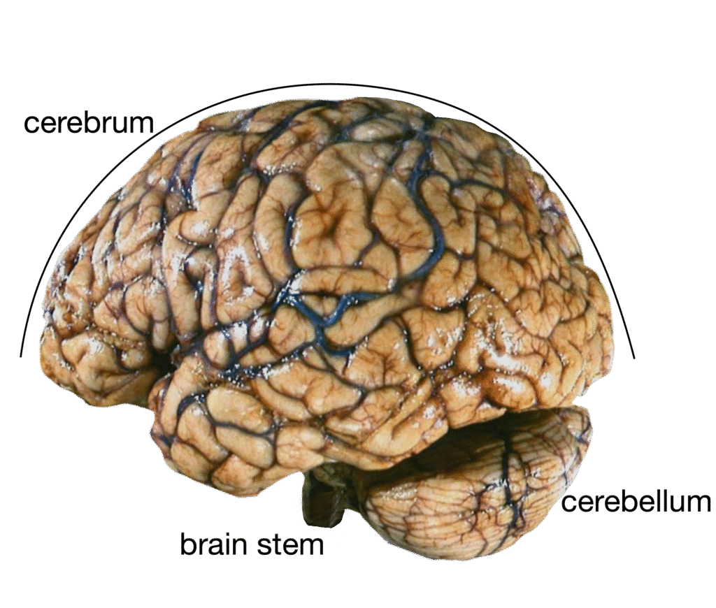
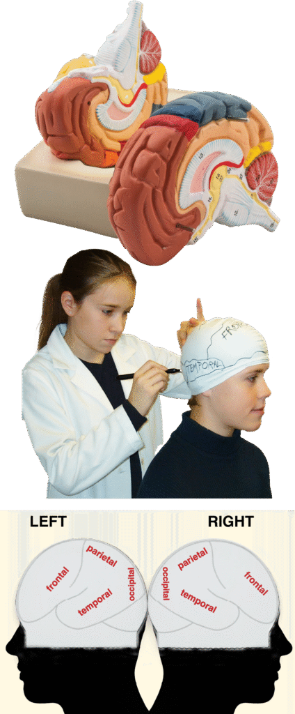
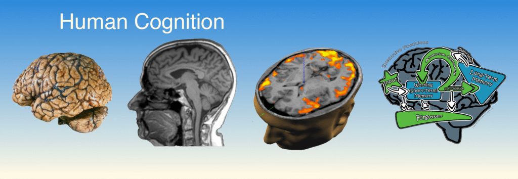
Enhance student understanding of the brain’s anatomy and its correlation with function.
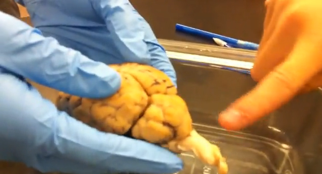
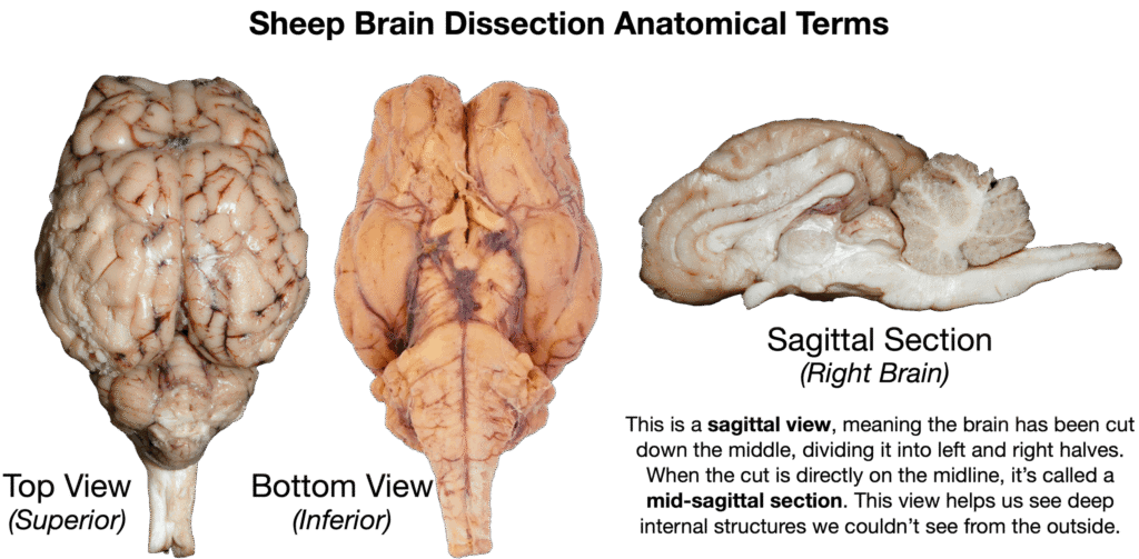
Develop proficiency in identifying and modeling major brain structures.
Foster skills in comparative anatomy through hands-on dissection and modeling activities.