
Back to: Human Cognition – Teacher
Student Portal
Investigation 2: Concepts
Welcome to the Concepts section of Investigation 2.
As you explore these slides, discover how your nervous system continuously receives information, processes it, and helps you respond to the world around you.
Before You Begin
These slides introduce the main ideas you’ll investigate in this lesson. As you work through them, look for examples of how the nervous system receives information, processes it, and produces responses.
Think Like a Scientist
As you explore the slides in this Investigation, think carefully about the questions that follow each slide. Your teacher may ask some or all of these questions during class discussion. Don’t worry about finding the “right” answer immediately—observe carefully, think critically, and be prepared to explain your reasoning.
Click on any image below to expand
SLIDE HC-2-1
Title: Human Cognition – Investigation 2
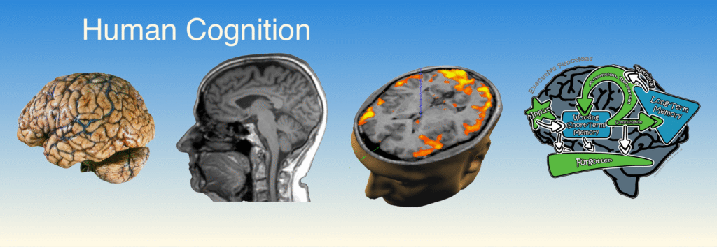
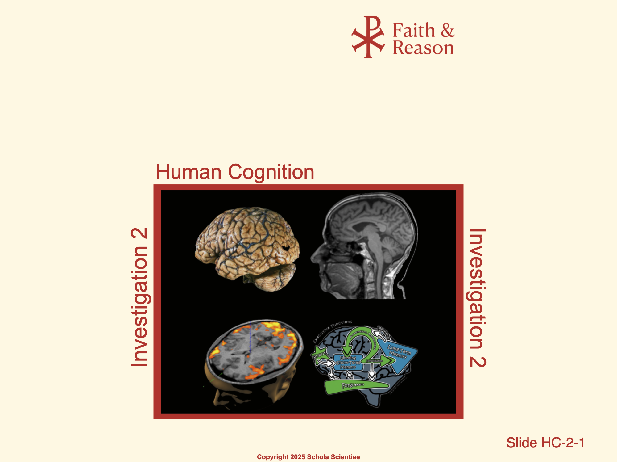
This slide introduces Investigation 2: Simple Information Processing. It reminds us that the brain is not only a physical organ but also the center of thought, memory, and decision-making. The images highlight several perspectives: a preserved brain specimen, a brain MRI, brain activity measured by fMRI, and a schematic of memory systems. Together, they emphasize that cognition is both biological and functional—what the brain is and what the brain does.
This Investigation will begin by exploring the structure of the brain and how it allows for increasingly complex forms of processing.
Discussion Questions and Answers:
- Q1: What does this slide show?
- Q2: Why do you think we are starting this unit with these images?
- Q3: What do you notice about the difference between the specimen, scan, and diagram?
- Q4: Why might the brain be the best place to start when studying human cognition?
SLIDE HC-2-2
Title: The Human Brain
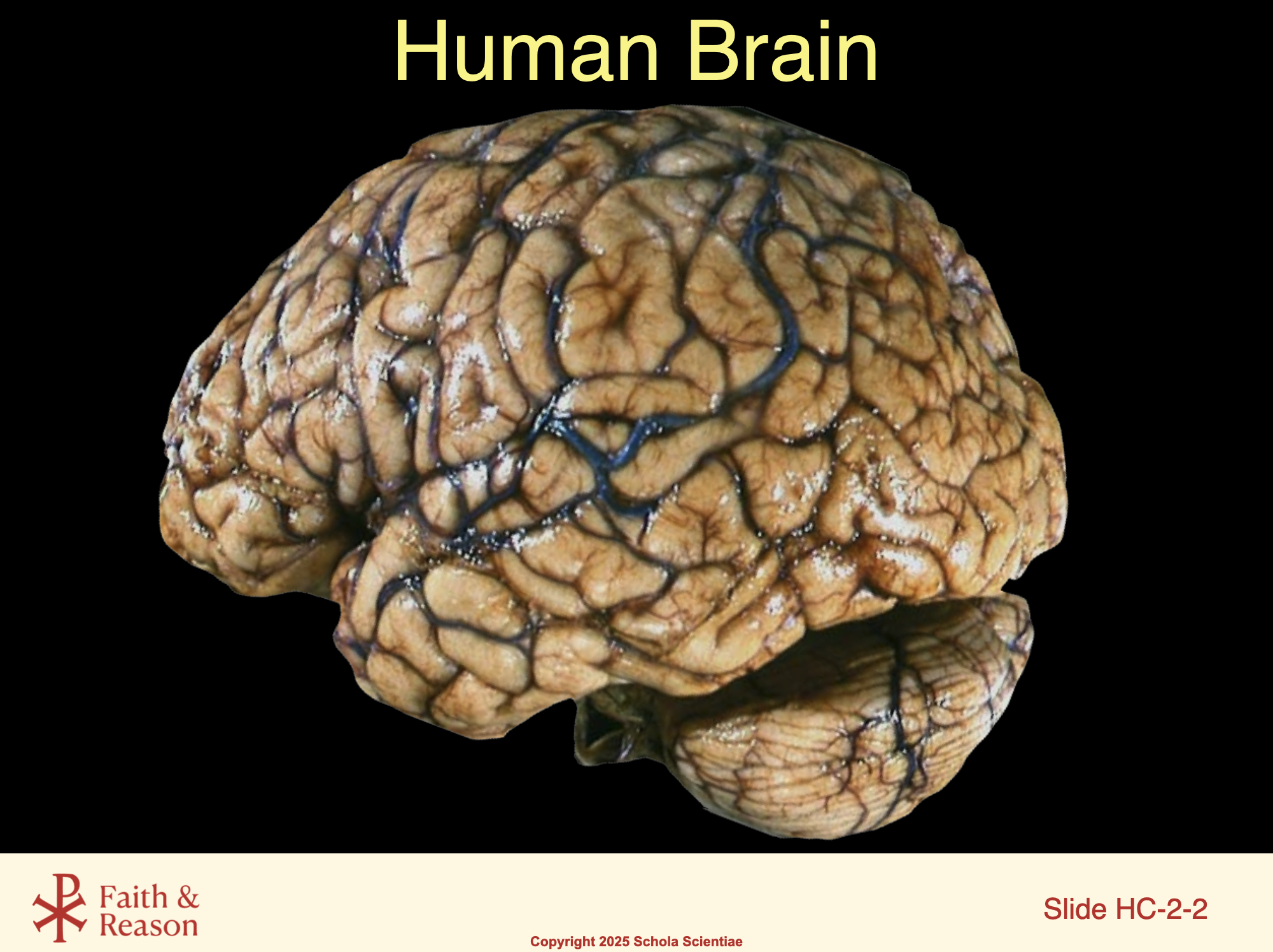
This image shows an actual human brain specimen. Notice its wrinkled surface, called the cerebral cortex, which provides increased surface area for billions of neurons. The folds allow for higher levels of information processing, from sensory perception to reasoning and planning. While the brain may look like a single organ, it is in fact divided into specialized regions that constantly communicate, allowing us to perform both simple reflexes and complex thought. In this investigation, we will examine how the brain’s physical design supports its extraordinary cognitive functions.
Discussion Questions and Answers:
- Q1: What does this photo show?
- Q2: Why do you think the brain has so many folds and grooves?
- Q3: How does this physical structure help explain human intelligence?
- Q4: What questions come to your mind when looking at a real human brain?
SLIDE HC-2-3
Title: Nervous
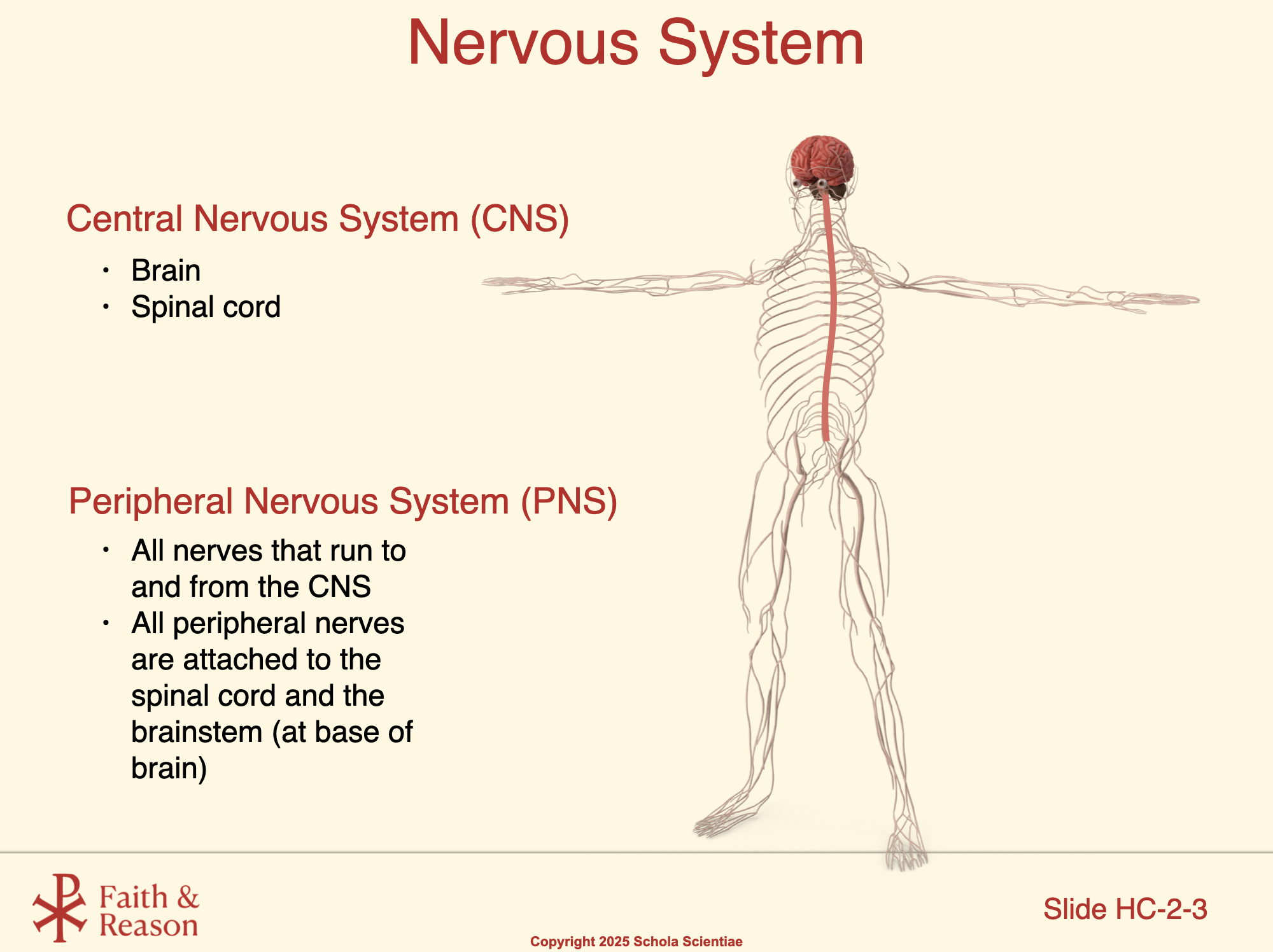
This slide introduces the two major divisions of the nervous system:
1. Central Nervous System (CNS)
- Consists of the brain and spinal cord.
- Acts as the body’s control center, where information is processed and decisions are made.
- The brain handles higher functions like memory, learning, and reasoning.
- The spinal cord coordinates simple, rapid responses like reflexes.
2. Peripheral Nervous System (PNS)
- Includes all nerves that run to and from the CNS.
- Nerves connect the CNS to the rest of the body — muscles, skin, and organs.
- All peripheral nerves are attached either to the spinal cord or to the brainstem at the base of the brain.
- Think of the PNS as the communication network that carries information back and forth between the CNS and the body.
Key Teaching Point:
This slide sets up the whole unit. Students should recognize that the CNS is the “command center,” while the PNS is the “messenger system” that links the body and the brain/spinal cord.
Discussion Questions and Answers:
- Q1: What are the two main parts of the Central Nervous System?
- Q2: What is the job of the CNS?
- Q3: What is included in the Peripheral Nervous System?
- Q4: Where are peripheral nerves attached?.
- Q5: How could you describe the difference between the CNS and the PNS?
SLIDE HC-2-4
Title: Information Processing
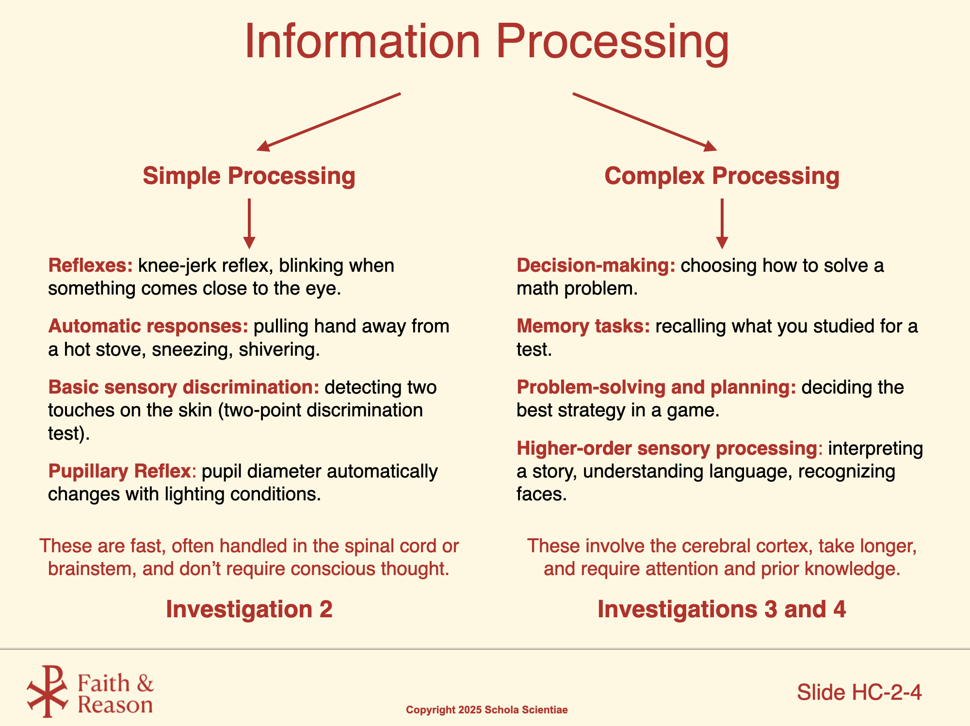
This slide introduces two categories of information processing in the nervous system: Simple Processing and Complex Processing.
Left Side – Simple Processing
Examples include reflexes like the knee-jerk reflex or blinking, automatic responses such as sneezing or pulling your hand away from a hot stove, pupil dilation reflex, and basic sensory discrimination like detecting two touches on the skin during the two-point discrimination test.
- Simple processing is fast, automatic, and often protects us by allowing rapid responses before conscious thought occurs.
- They don’t require conscious thought.
- This will be the focus of Investigation 2, where students will study reflexes and basic neurological exams.
Right Side – Complex Processing
Examples include decision-making (choosing how to solve a math problem), memory tasks (recalling what you studied), problem-solving and planning (deciding a strategy in a game), and higher-order sensory processing (interpreting a story, understanding language, recognizing faces).
- These involve the cerebral cortex, require more time, attention, and prior knowledge.
- This type of processing will be the subject of Investigations 3 and 4, where students will explore memory, learning, and higher-level cognition.
Key Contrast for Students:
Point out how simple processing keeps us safe through immediate, automatic responses, while complex processing allows us to think, learn, and plan at a higher level.
This distinction sets the stage for the rest of the Human Cognition Unit, moving from quick, protective responses in Investigation 2 to the deeper functions of the brain in Investigations 3 and 4.
Discussion Questions and Answers:
- Q1: What is the main difference between simple and complex information processing?
- Q2: Can you give an example of simple processing?
- Q3: What about an example of complex processing?
- Q4: Which type of processing will we focus on in Investigation 2?
- Q5: Which type of processing will we study in Investigations 3 and 4?
SLIDE HC-2-5
Title: The Reflex Arc (Knee-Jerk Reflex)
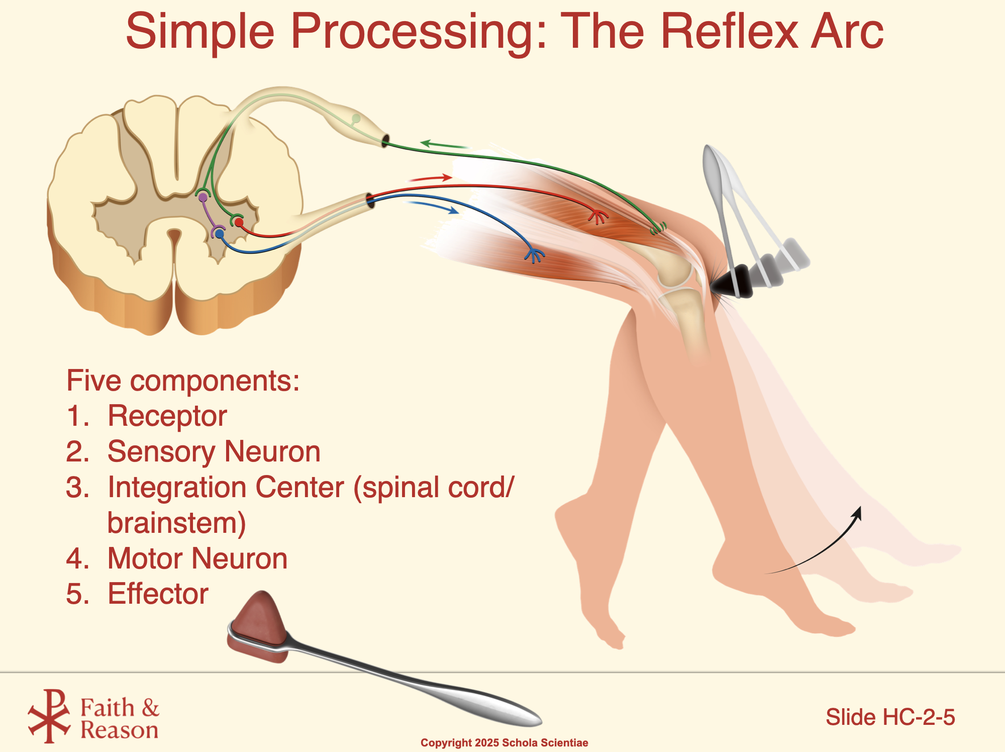
This slide shows the classic patellar (knee-jerk) reflex, an example of simple information processing. We can identify each of the five components of the reflex arc:
- Receptor: The muscle spindle in the quadriceps detects when the tendon is tapped and stretched.
- Sensory Neuron: The stretch signal travels from the receptor to the spinal cord through a sensory neuron (green).
- Integration Center (Spinal Cord): In the spinal cord, the sensory neuron synapses directly with a motor neuron (red) — this is a monosynaptic reflex. At the same time, an interneuron (purple) sends an inhibitory signal to the neuron (blue) that normally activates the hamstrings.
- Motor Neuron: The motor neuron (red) sends the return signal back to the quadriceps muscle.
- Effector: The quadriceps muscle contracts, producing the lower leg “kick.”
- Inhibitory Effect on the Hamstrings: To make the movement smooth, the spinal cord also inhibits the hamstrings (blue neuron). This is called reciprocal inhibition — while the quadriceps contract, the hamstrings relax, preventing them from resisting the motion.
👉 Teacher Note: Highlight how this reflex never needs to “wait” for the brain. The signal loop stays within the spinal cord, allowing the body to respond before conscious awareness occurs. The nervous system values speed when protection is more important than analysis. Then connect to the larger point: this is an example of simple processing in the nervous system.
Discussion Questions and Answers:
- Q1: What part of the body detects the stretch when the knee is tapped?
- Q2: Where does the sensory signal travel first?
- Q3: Does the brain have to process this signal before the leg kicks?
- Q4: Which muscle contracts to cause the kick?
- Q5: Why don’t the hamstrings resist this movement?
SLIDE HC-2-6
Title: Other Examples of Simple Processing
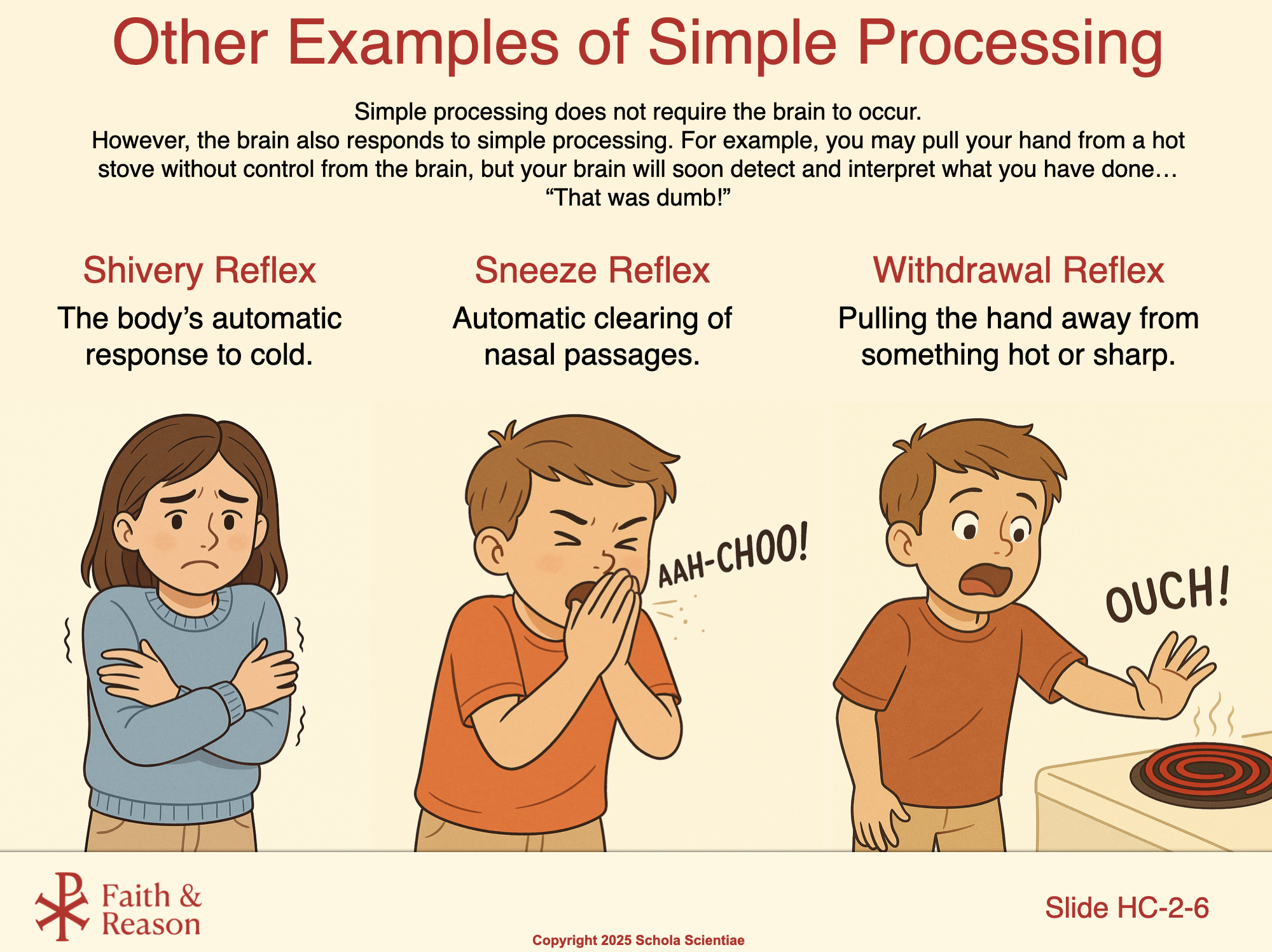
This slide shows that not all reflexes look the same, but they all share the same feature: they are automatic, protective, and fast.
- Shivery Reflex: The body’s automatic response to cold. Muscles contract rapidly to generate heat without conscious control.
- Sneeze Reflex: A powerful, automatic clearing of the nasal passages. This protects the lungs from dust or irritants.
- Withdrawal Reflex: Pulling the hand away from something hot or sharp. The spinal cord directs the action before the brain is even aware of the danger.
Key Idea:
Reflexes happen so quickly because the spinal cord and brainstem can respond before you become consciously aware of the stimulus. Your brain then interprets what happened, helping you understand the event and decide what to do next. This partnership between automatic responses and conscious thought is an example of simple information processing.
Discussion Questions and Answers:
- Q1: What is the purpose of shivering when we are cold?
- Q2: Why is sneezing considered a reflex?
- Q3: What makes the withdrawal reflex so important?
- Q4: If the spinal cord can make your hand pull back from a hot stove, what role does the brain play?
- Q5: How are all of these reflexes similar?
SLIDE HC-2-7
Title: Skin Receptors
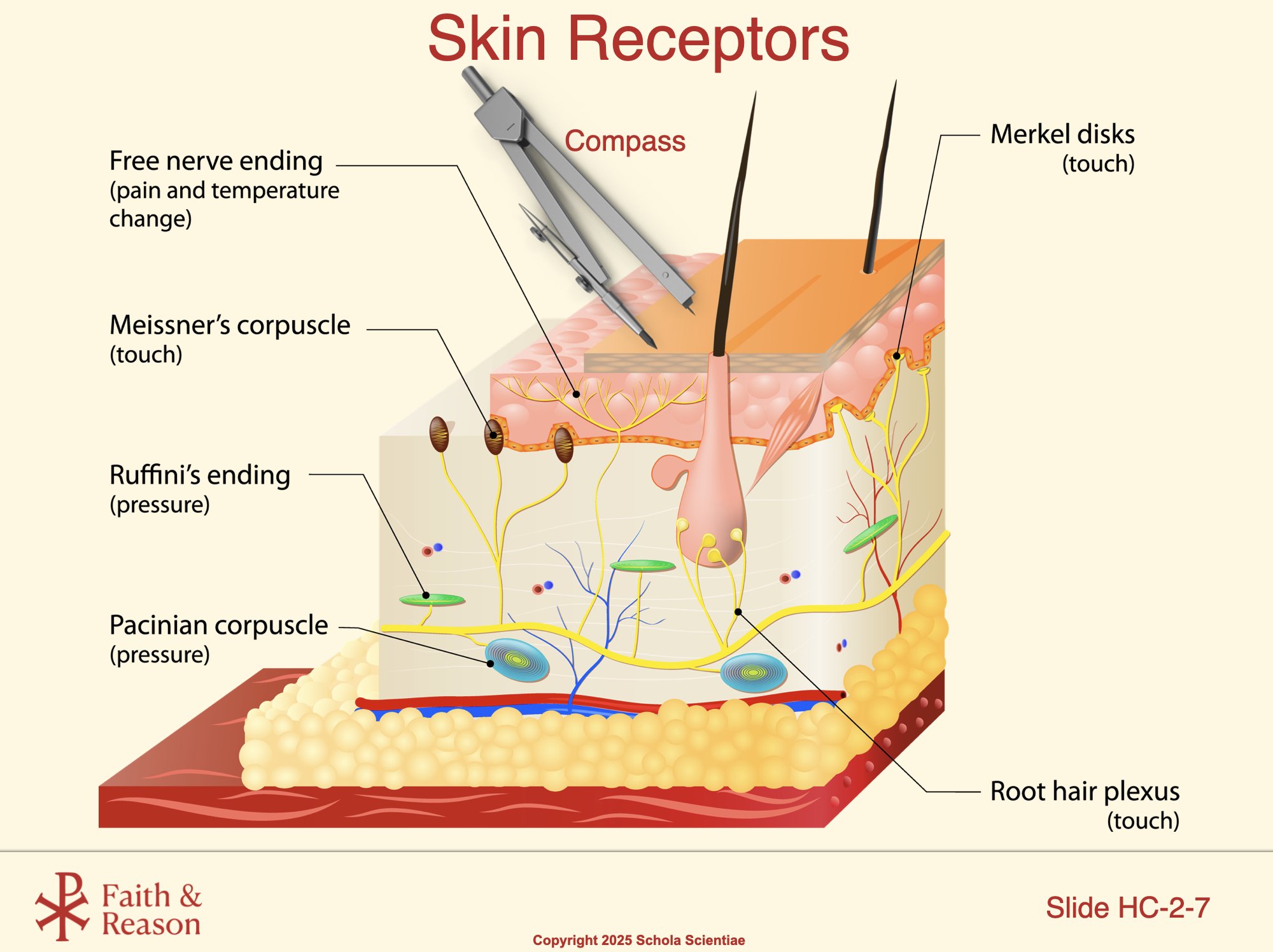
This slide shows the different types of receptors in the skin that allow us to sense our environment. Notice that there are many kinds, each tuned to different signals:
- Free nerve endings
- Merkel disks
- Meissner’s corpuscles
- Ruffini endings
- Pacinian corpuscles
- Root hair plexus
You don’t need to go into detail about each one here — the main point is that the skin contains a variety of specialized nerve endings.
Also notice the compass at the top of the slide. Students will use a compass in Investigation 2 to perform the two-point discrimination test. This lab will help them see that receptor density is different across body regions, which explains why some areas (like fingertips) are more sensitive than others (like the back).
Key Idea: The skin is filled with specialized receptors, and we’ll use the compass to explore how their distribution affects sensitivity.
Discussion Questions and Answers:
- Q1: What do all the receptors shown in the skin diagram have in common?
- Q2: Why will we be using a compass in the lab?
- Q3: Why are fingertips more sensitive than the back?
- Q4: Why might some parts of the body need more detailed information about touch than others?
SLIDE HC-2-8
Title: Sensory Homunculus
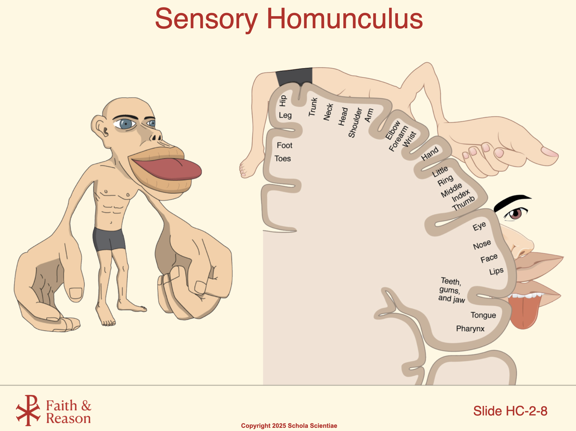
This slide shows the sensory homunculus, a map of how much brain area in the sensory cortex is devoted to different parts of the body.
Notice that the homunculus figure looks distorted: the hands, lips, and tongue appear very large compared to the arms, legs, or back.
This distortion shows that areas with high receptor density (lots of nerve endings, like fingertips and lips) take up more space in the brain.
Areas with low receptor density (like the back or thighs) take up much less space.
Key Idea: Sensitivity doesn’t match body size — it matches the importance of the body part for survival and interaction. Hands are crucial for tool use, lips and tongue for eating and speech, so they are represented as oversized.
This slide connects directly to the two-point discrimination test in Investigation 2. Students will discover that what they experience in the lab matches the homunculus map of the brain.
Discussion Questions and Answers:
- Q1: Why does the homunculus have such big lips and hands?
- Q2: Why are the arms, legs, and back much smaller in the homunculus?
- Q3: What is the main message of the homunculus?
- Q4: How does this connect to the two-point discrimination test you’ll do in lab?
- Q5: What does the homunculus tell us about the way the brain processes information?
- Q6: If the brain gives more attention to what matters most for survival and interaction, what kinds of things deserve our attention in everyday life?
SLIDE HC-2-9
Title: Pupillary Reflex
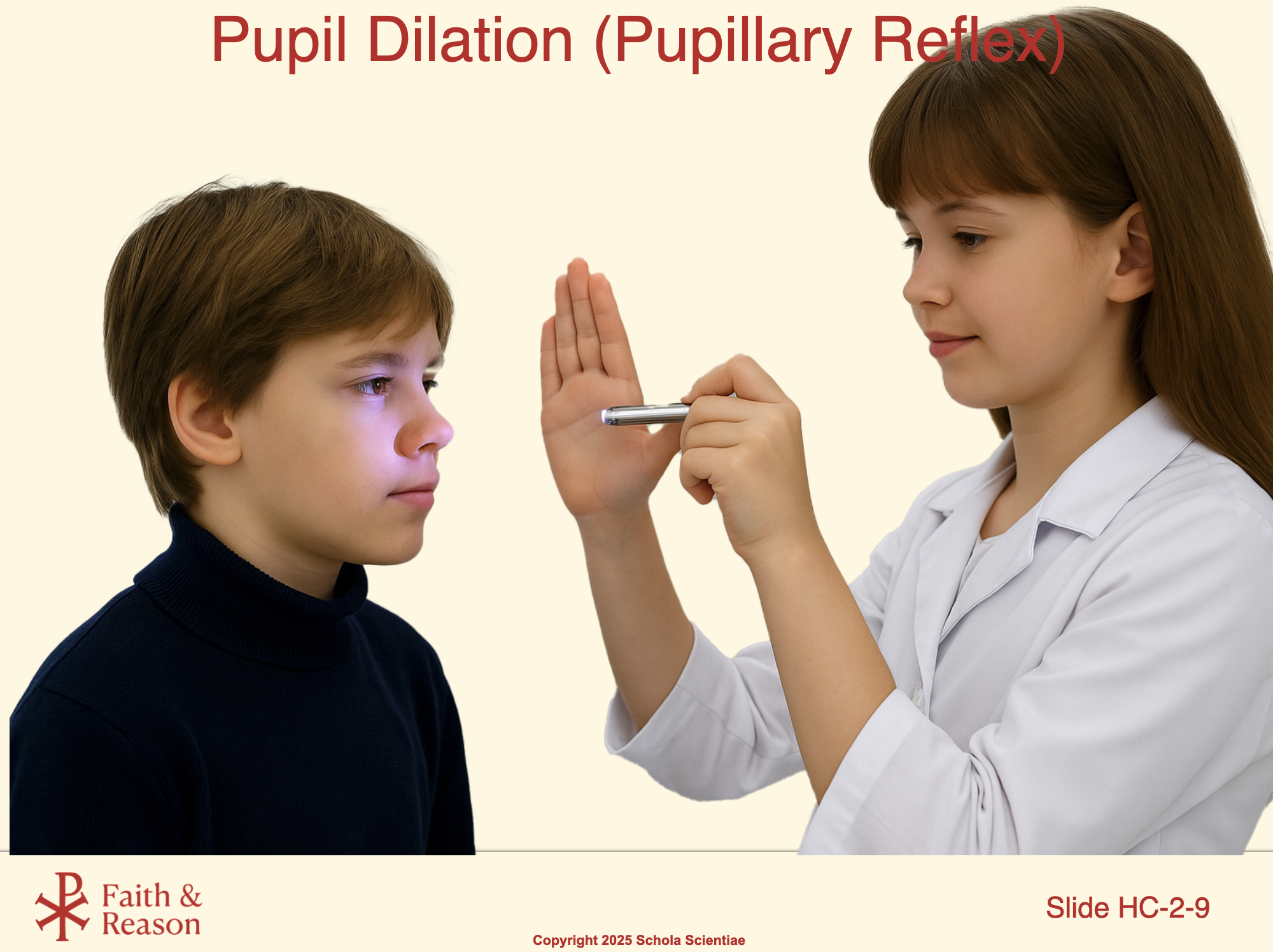
This slide shows the pupillary light reflex, another example of a simple, automatic processing pathway.
When light shines into the eye, the pupil constricts to limit how much light enters.
In darkness, the pupil dilates to let in more light.
This reflex happens automatically and very quickly to protect the eye and help vision adjust to changing light.
Key Point: Even though it feels automatic, the pathway still involves both the peripheral nervous system (the sensory input from the eye and the motor signal to the iris muscles) and the central nervous system (the signal passes through the brainstem where it is processed before the motor command is sent back).
In the Lab: Students will perform this reflex test in Investigation 2 using a small flashlight, just as doctors do in a routine eye exam. They’ll observe the pupil’s quick response to light and notice how the reflex does not require conscious thought.
Discussion Questions and Answers:
- Q1: What happens to the pupil when light shines into the eye?
- Q2: Which parts of the nervous system are involved in this reflex?
- Q3: Why is this reflex important for vision?
- Q4: How will students test this reflex in the lab?
- Q5: Why would it be dangerous if the pupil did not respond automatically to light?
SLIDE HC-2-10
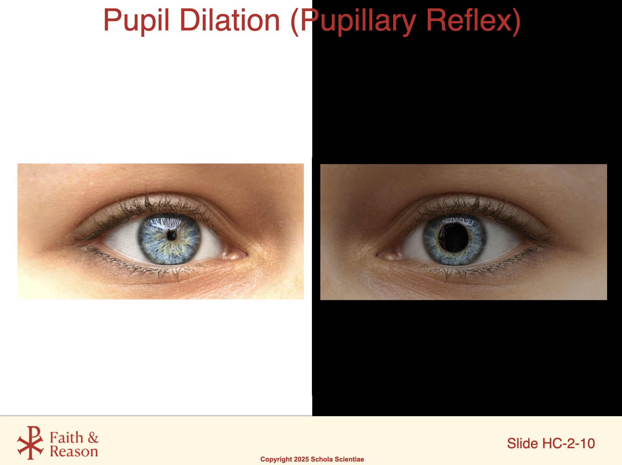
This slide shows how the pupils respond automatically to changes in light. In bright light (left), the pupil constricts to limit the amount of light entering the eye. In dim light (right), the pupil dilates to allow more light in. This is an example of an involuntary reflex controlled by the autonomic nervous system. The response travels from the eye to the brain and back, protecting vision and ensuring that the right amount of light reaches the retina.
Discussion Question and Answer:
- Q1: Why would it be difficult to function if your pupils could not change size?
SLIDE HC-2-11
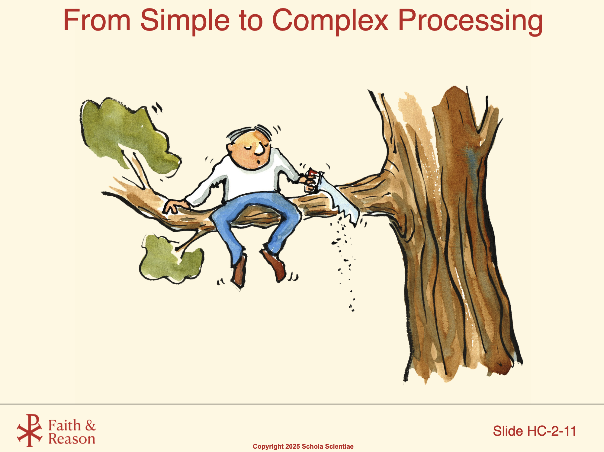
This cartoon is a great reminder that reflexes can protect us in the short term, but they don’t replace critical thinking. Cutting off the branch you’re sitting on is not something a reflex can fix — it’s a mistake of poor planning. That’s why the human brain’s ability for complex processing — problem-solving, memory, reasoning — is so important. Investigations 3, 4, and 5 will help us explore those higher-level abilities.
Discussion Questions and Answers:
- Q1: Why wouldn’t a reflex save this person?
- Q2: What kind of processing is needed to avoid this mistake?
- Q3: Why is complex processing just as important as reflexes?