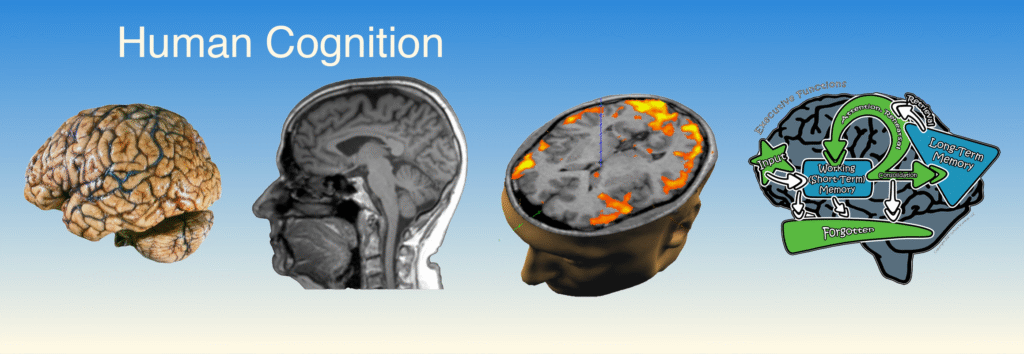
Explore the anatomy of the brain and how its structure relates to function.
Identify and model major brain structures.
Compare human and sheep brain anatomy through hands-on dissection and modeling activities.
As you work: Treat the sheep brain as scientific evidence, not simply as a specimen to identify. Compare what you observe with the human brain model and think about how each structure relates to its function.
Be Prepared
For this lab, your group will need the following materials:
Chromebook/Laptop with Student Guide
Human brain model
Whole sheep brain
Sectioned sheep brain
Dissection pads and probes
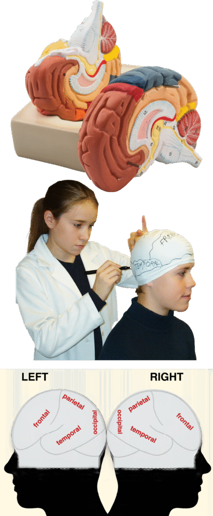
Swim cap (1 per student)
Markers
Lab Reminder: Handle all biological specimens and dissection tools carefully and follow your teacher’s safety instructions. Work slowly and observe carefully—the goal is not to finish quickly, but to make accurate observations.
Investigate
Sheep Brain Dissection:
In this Investigation, you will examine the structure of a sheep brain and compare what you observe with the human brain structures you have studied. You will then use a brain cap model to explore the location and functions of the major cerebral lobes.
As you work, observe carefully and think about the relationship between structure and function. Record your observations in your Student Guide and be prepared to explain what your observations suggest about how the brain is organized.
Observation Reminder: Record what you actually observe, even if it differs from what you expected to see.
Experiment
This experiment is made up of two parts:
Sheep brain dissection
Brain cap modeling activity
Below is the step-by-step procedure you will follow for both parts of the lab:
PART 1: Sheep Brain Dissection
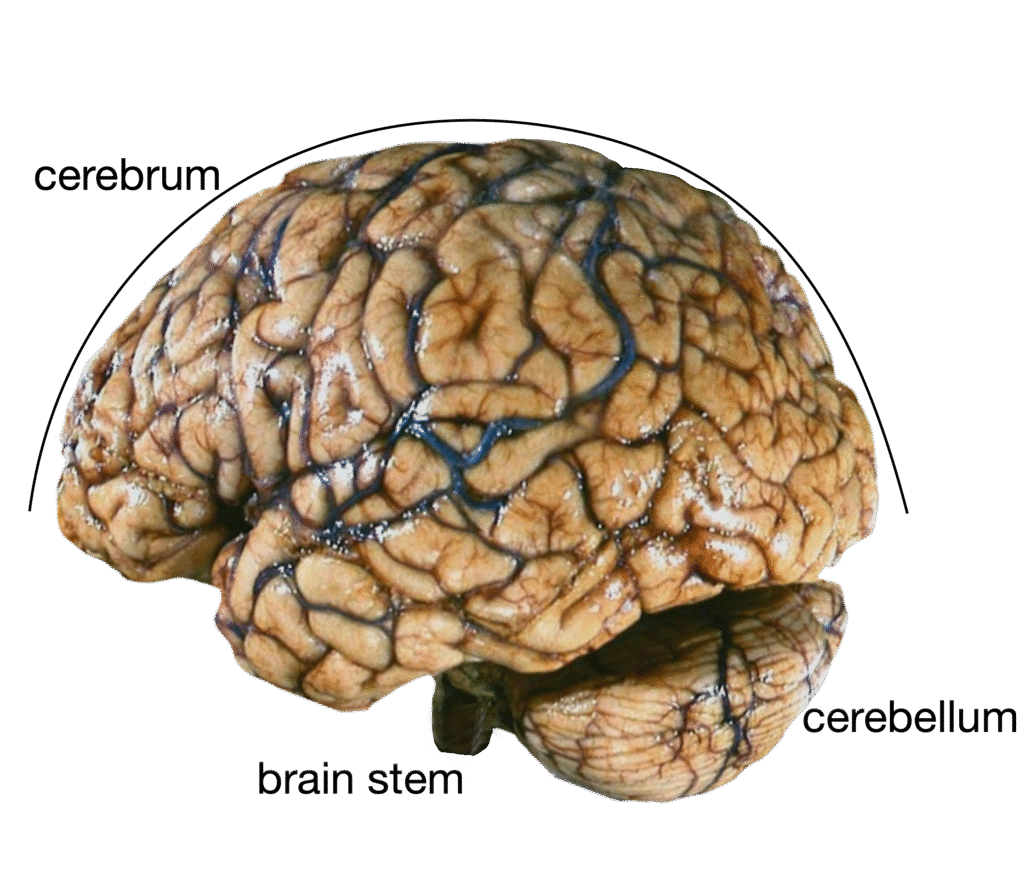
1. Observe the human brain model
Identify the cerebrum, cerebellum, and brainstem.
Note the wrinkled appearance caused by gyri (ridges) and sulci (grooves).
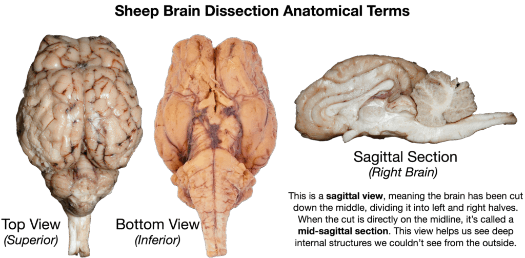
2. Examine the whole sheep brain
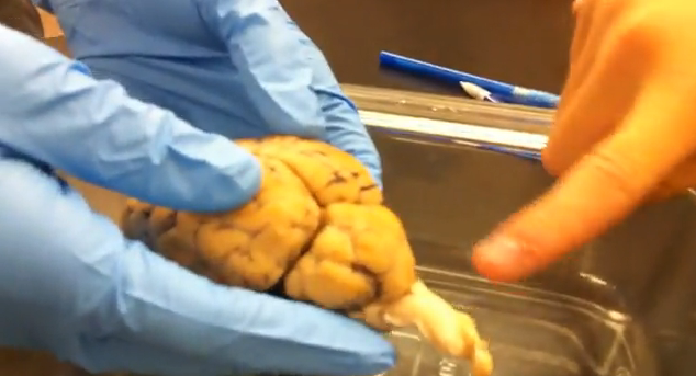
Locate and compare the cerebrum, cerebellum, and brainstem.
3. Use the dissection probe
Gently explore the surface to find gyri and sulci.
Identify the cerebral cortex — the outer layer of the cerebrum.
4. Compare the sheep and human brains
Discuss similarities and differences in shape, size, and orientation.
Pay special attention to the brainstem’s angle and the relative size of the cerebellum.
PART 2: Brain Cap Modeling Activity
1. Select one person in your group to wear the swim cap.
2. Trace brain features on the cap using a marker. a. Place the swim cap securely on your group member’s head. b. Have a partner outline the cerebral hemispheres and the cerebellum. c. Be sure to reverse the orientation when labeling the opposite side.
3. Add major anatomical landmarks to the cap:
Longitudinal fissure – groove dividing left and right hemispheres
Central sulcus – vertical groove near the top center
Lateral sulcus – horizontal groove near the side of the brain
4. Label the four major lobes:
Frontal lobe (front)
Parietal lobe (top)
Temporal lobe (side)
Occipital lobe (back)
5. Wear and present your labeled brain model.
Use the cap as a wearable study tool during discussion or review.