
Investigation 1: Concepts
NAVIGATE
Once the slide presentation is launched
- use your left and right arrows to advance or go back in the slide presentation, and
- hover your mouse over the left edge of the presentation to get a view of the thumbnails for all the slides so that you can quickly move anywhere in the presentation.
Click on any image below to expand
SLIDE HC-1-1
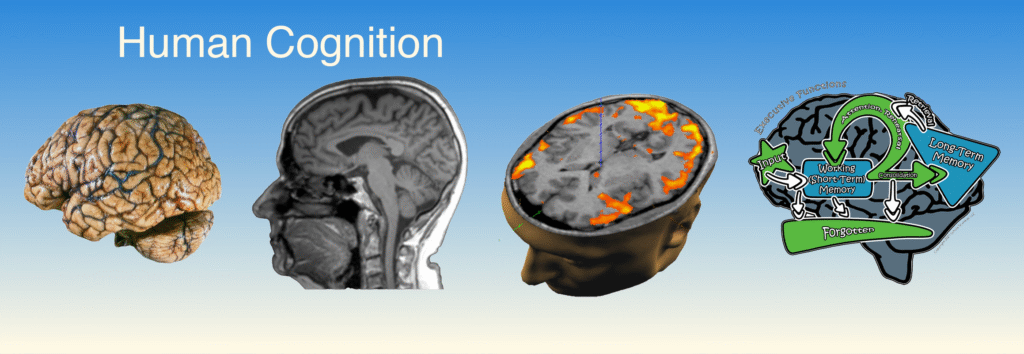
Title: Human Cognition – Investigation 1
Welcome to Investigation 1 of our Human Cognition unit. In this first investigation, we begin to explore one of the most complex and mysterious systems in the natural world: the human brain.
Throughout this unit, we’ll examine how your brain takes in information, makes decisions, stores memories, and helps you become the person you are.
In this investigation, we’ll focus on:
Basic structure and functions of the brain
Major brain regions and what they control
How we first discovered that different brain areas have different purposes
The beginning of our journey through the Information Processing Model—your brain’s inner operating system
We’ll also look at a fascinating historical case: Phineas Gage, whose injury gave scientists the first real clue about how the brain affects behavior and personality.
This investigation includes two hands-on lab activities:
A brain dissection, where we’ll compare sheep and human brains
A brain cap activity, to map the major lobes of the brain on your own head!
Discussion Questions and Answers:
Q1: What will we be learning in this first investigation?
A1: We’ll learn about the brain’s structure and major functions, and how scientists started understanding what different brain parts do.
Q2: Who was Phineas Gage, and why is he important in this unit?
A2: He was a railroad worker who survived a serious brain injury. His case showed that brain injuries can change personality, helping scientists link brain regions to behavior.
Q3: What are the two lab activities we’ll be doing?
A3: A brain dissection and a brain cap exercise—both will help us see and understand how the brain is organized.
SLIDE HC-1-2
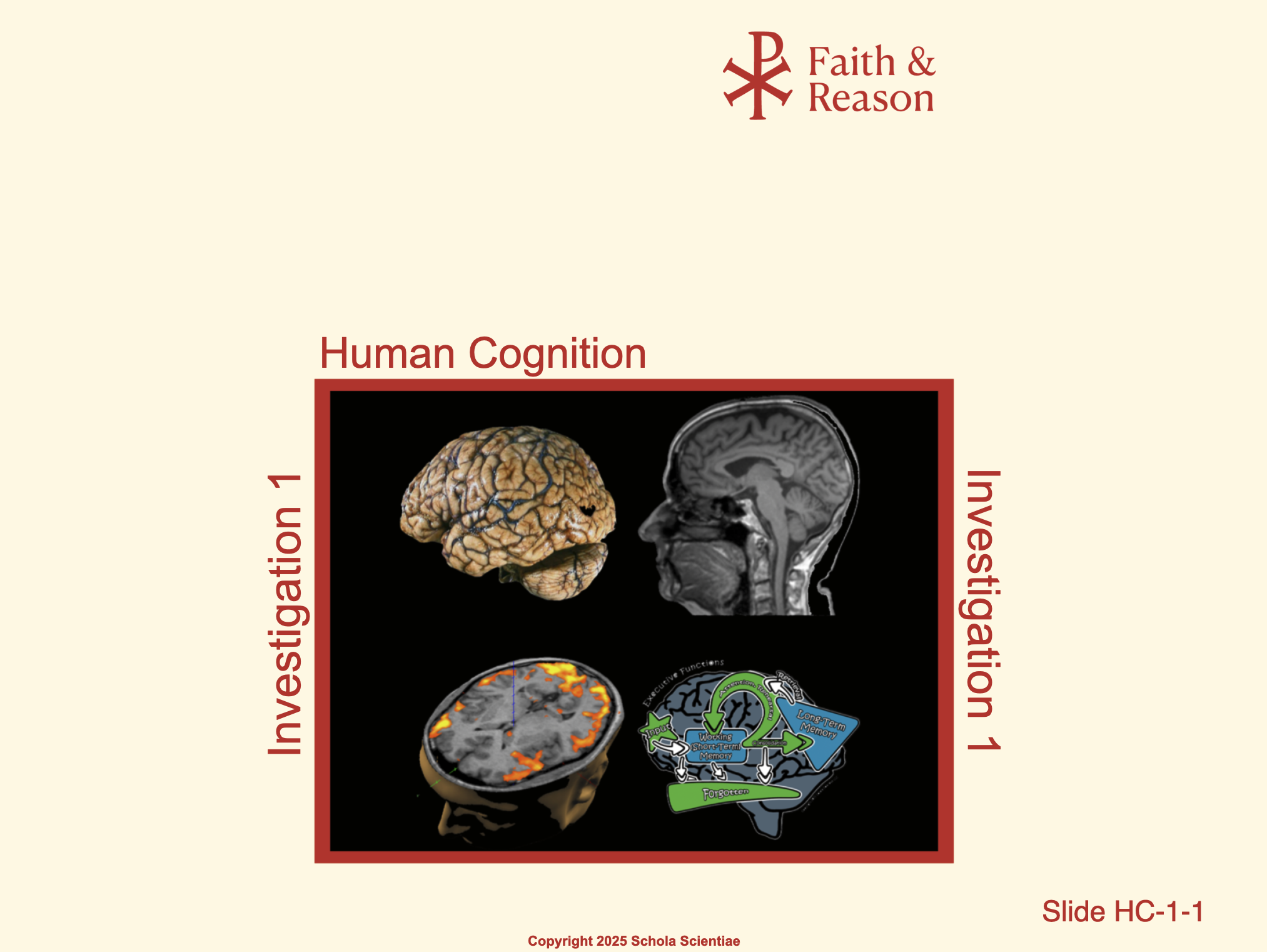
The Human Brain (Prenatal Development Overview)
This slide integrates several important parameters of human prenatal development into a single overview. It illustrates fetal growth by trimester, stages of maternal pregnancy, and the increasing chances of survival outside the womb as gestation progresses. The graphic also ties brain development to these stages, emphasizing that neurological growth begins in the first trimester and continues through adolescence.
Students who have already completed the Human Prenatal Development unit will recognize this slide, making it a familiar anchor point. Using it here helps tie the Human Cognition unit back to prior knowledge, reinforcing continuity across the two programs and preparing students to connect early brain development with later cognitive functions.
Discussion Questions and Answers:
Q1: What does this slide show?
A1: It shows how a baby develops during pregnancy, including growth in each trimester, the mother’s changes, and the chances of survival outside the womb.
Q2: Why is this slide important in our study of the brain?
A2: It reminds us that brain development begins very early in pregnancy and continues for many years after birth, which connects to how we study human cognition.
Q3: Why do you think we are reusing this slide from the Human Prenatal Development unit?
A3: To connect what students may already know about prenatal growth with this new unit on how the brain works throughout life.
SLIDE HC-1-3
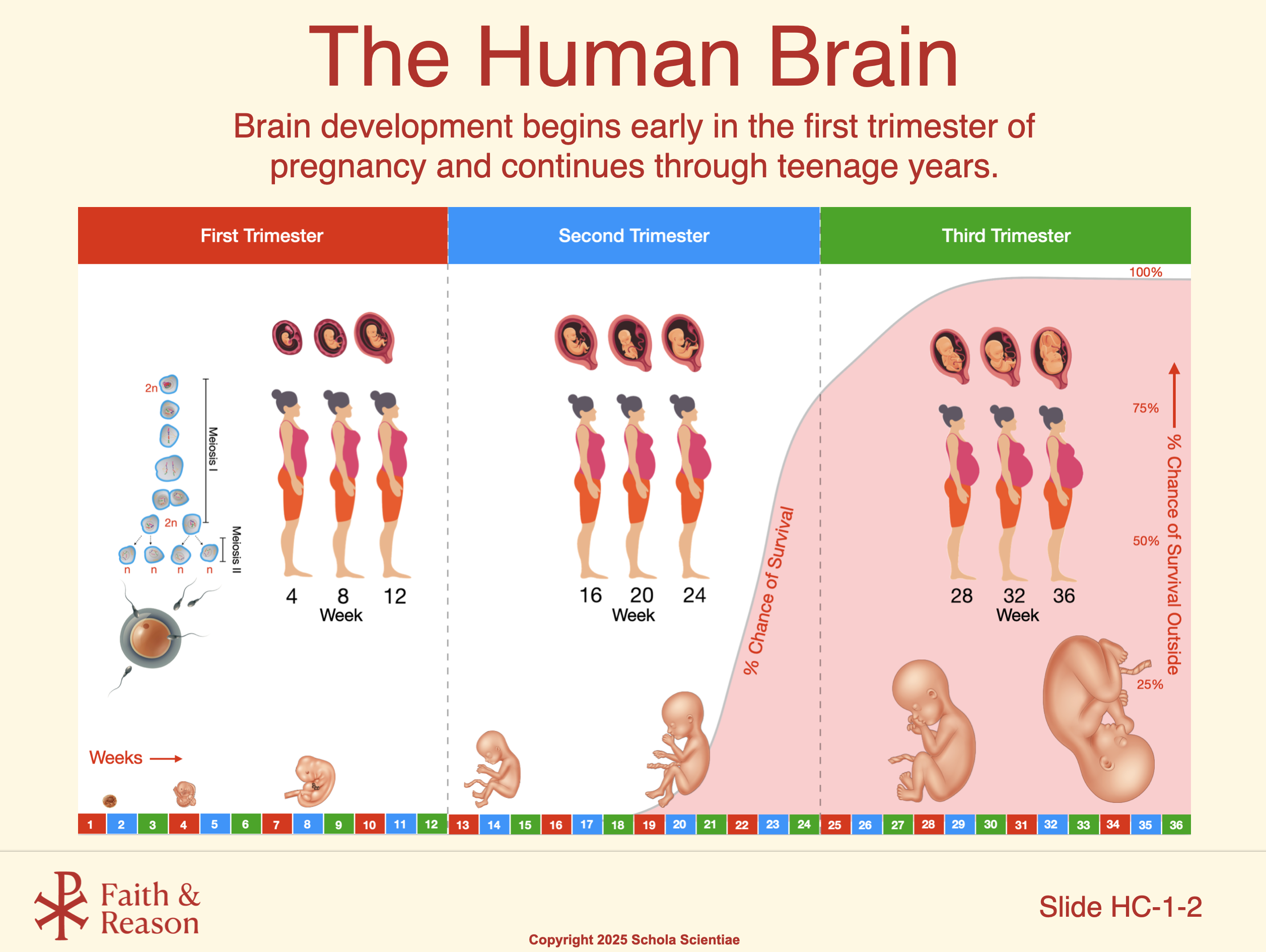
The Human Brain (Early Growth in Detail)
This slide zooms in on the developing brain itself, showing dramatic changes in size and structure from just under a month after conception through birth. At five weeks, specialized brain regions begin to form, marking the start of complex neural organization. By eight weeks, the brain is producing an astonishing 250,000 neurons per minute, a pace that underscores the intensity of prenatal brain growth.
Together with the previous overview slide, this image helps students appreciate both the broad timeline of brain development and the microscopic pace of neuron formation. For those who studied the Human Prenatal Development unit, these details reinforce earlier lessons and prepare students to connect prenatal growth with the larger theme of cognition that unfolds throughout life.
Discussion Questions and Answers:
Q1: What does this slide show?
A1: It shows the developing brain at different stages, from about one month after conception through nine months.
Q2: Why is this important for our study of the brain?
A2: It shows how quickly the brain forms—specialized areas start appearing by five weeks, and by eight weeks the brain is making 250,000 new neurons every minute.
Q3: What does this tell us about the importance of the early stages of life?
A3: It shows that the brain’s foundation is built very early, so what happens before birth has a big impact on later development and cognition.
SLIDE HC-1-4
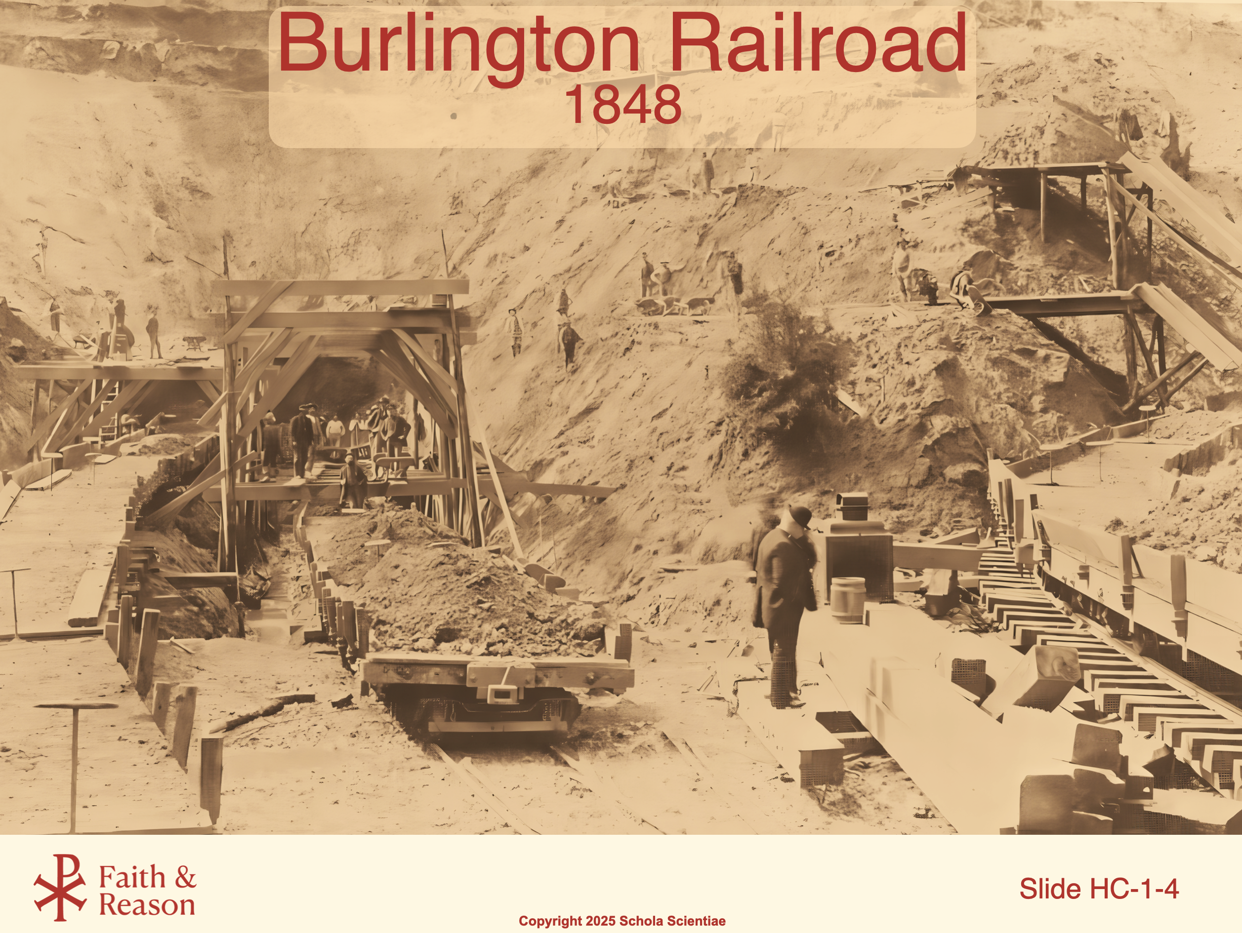
Title: Burlington Railroad – 1848
This photograph shows workers digging a railroad cut in Vermont in the year 1848. The construction site is part of the Burlington Railroad, one of the largest and most important railroads in the early United States.
You might be wondering—what does this have to do with the brain?
This is the exact railroad where Phineas Gage was working when a terrible accident changed both his life and the field of neuroscience forever. Gage was using explosives to clear rocks for this railroad when a metal rod was accidentally driven through his skull.
This photo gives us historical context. It helps us imagine the world Phineas lived in, the kind of work he did, and how dangerous his job really was.
We’ll come back to this railroad throughout the investigation, because it’s where one of the most important early clues about the function of the frontal lobe came from.
Discussion Questions and Answers:
Q1: What does this photo show?
A1: It shows a railroad construction site from 1848—the same time and place where Phineas Gage was injured.
Q2: Why is this railroad important in our study of the brain?
A2: It’s where Phineas Gage had his accident, which helped scientists learn that different brain areas control different things.
Q3: Why do you think scientists still talk about a railroad worker from 1848?
A3: Because his injury revealed that damage to the brain can change someone’s behavior and personality—a major discovery at the time.
SLIDE HC-1-5
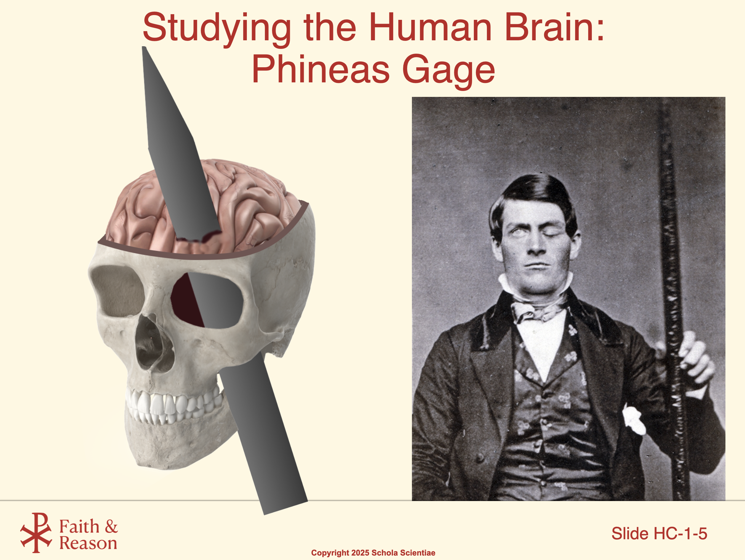
Title: Phineas Gage
This is Phineas Gage, a 25-year-old railroad foreman who survived one of the most famous accidents in medical history.
While working with explosives, a long iron rod was blasted straight through his skull and brain, entering through his cheek and exiting through the top of his head. Amazingly, he survived—but he was never the same.
Before the accident, Gage was calm, responsible, and well-liked. Afterward, he became rude, impulsive, disrespectful, and unreliable. This dramatic change in personality shocked doctors and his friends.
At the time, scientists didn’t understand how different parts of the brain controlled different behaviors. But Gage’s case gave them evidence that the brain is not just one big thinking blob—it has specialized areas with different jobs.
We’re still learning from Phineas Gage today. His story is a powerful example of how science often begins with unexpected observations.
Discussion Questions and Answers:
Q1: What happened to Phineas Gage?
A1: A metal rod shot through his skull during a railroad accident—he survived but his personality changed dramatically.
Q2: Why was his case important to science?
A2: It showed that damage to certain parts of the brain—like the frontal lobe—can affect emotions, behavior, and decision-making.
Q3: What does this teach us about the brain?
A3: That different regions of the brain control different functions—like thinking, emotions, and self-control.
SLIDE HC-1-6
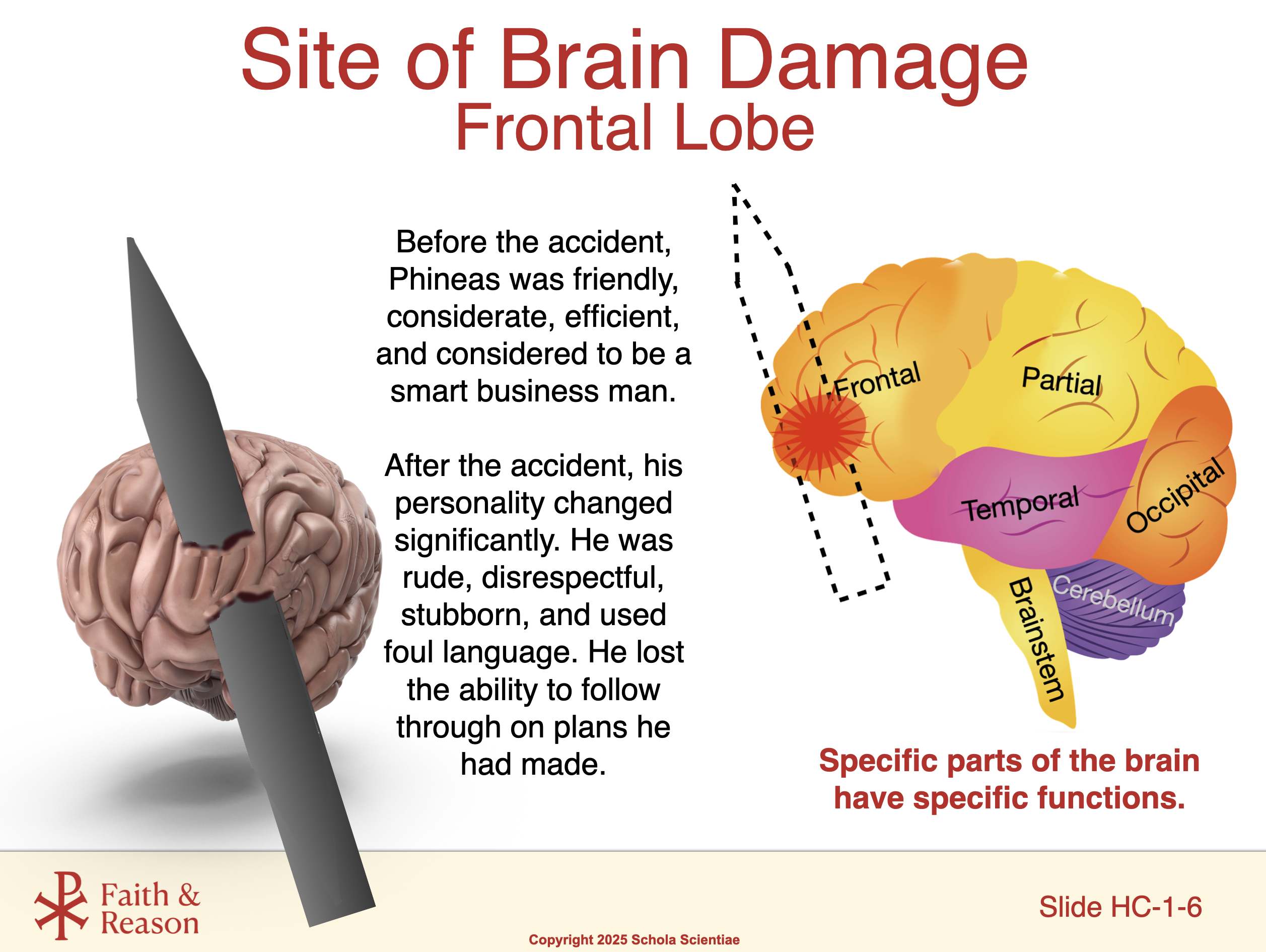
Title: Site of Brain Damage
This image shows a modern brain scan with a highlighted area—this is the frontal lobe, the region that was damaged in Phineas Gage’s accident.
The frontal lobe is located just behind your forehead. It helps control your personality, decision-making, problem-solving, and ability to control impulses. When this part of Phineas’s brain was destroyed, it didn’t affect his ability to move or speak—but it completely changed how he behaved.
Before Gage, people thought the brain was like a general-purpose machine. His case helped scientists realize that different areas of the brain control different functions.
This is one of the first known examples of localization of brain function—the idea that specific parts of the brain have specific jobs.
Discussion Questions and Answers:
Q1: What part of Phineas Gage’s brain was damaged?
A1: The frontal lobe, which is responsible for personality, decision-making, and self-control.
Q2: What happened to him after that part was damaged?
A2: He survived, but his behavior and personality changed a lot—he became impulsive and irresponsible.
Q3: Why was this discovery important?
A3: It showed that different parts of the brain have different roles, and damage to a specific area can change how a person thinks or acts.
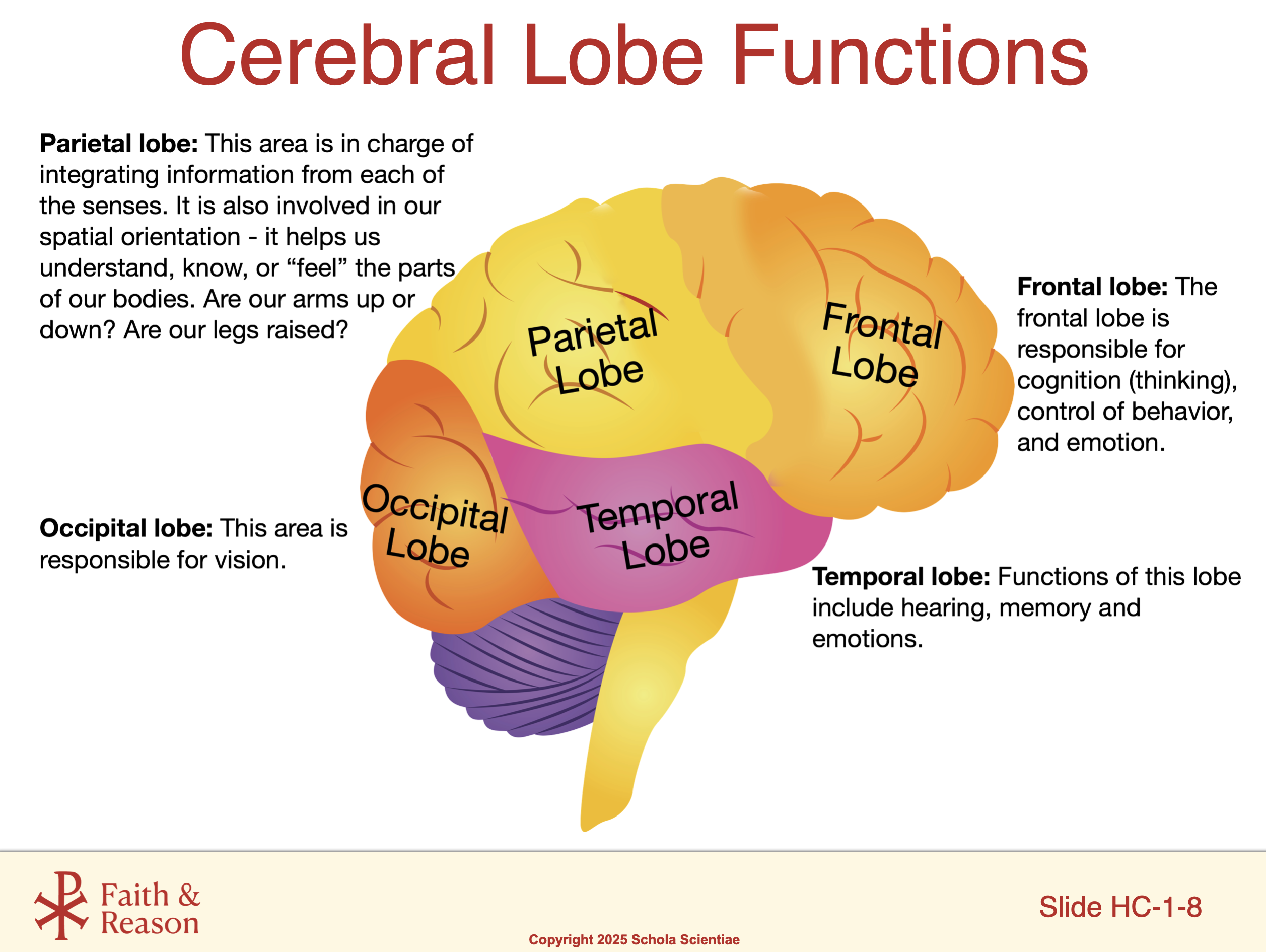
SLIDE HC-1-7
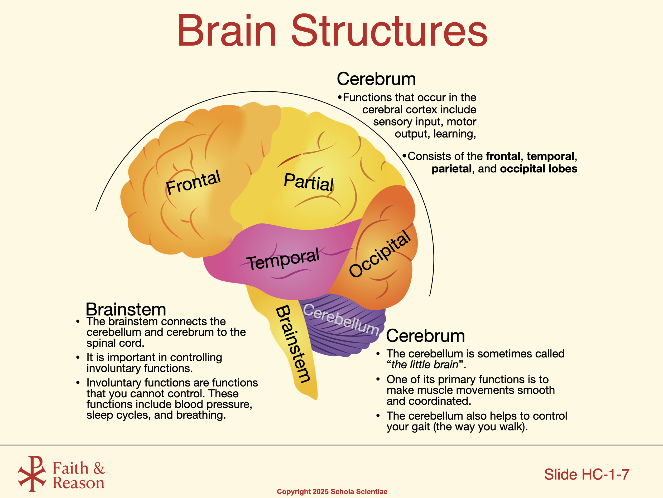
Title: Frontal Lobe and Behavior
This diagram shows the major components of the brain. Importantly, it indicates the location of the frontal lobe – the part of your brain that helps manage your thoughts, actions, and emotions. This is the part of the brain that was damaged in the case of Phineas Gage.
The frontal lobe is like the “executive” part of your brain—it helps you plan ahead, stay focused, resist distractions, and make wise choices. It’s also involved in controlling your emotions and helping you respond to situations in socially appropriate ways.
When the frontal lobe is healthy and active, it helps you act with self-control and think through your decisions. But when it’s damaged—like in the case of Phineas Gage—those abilities can weaken or disappear.
This slide shows that the structure of the brain is directly connected to behavior. It’s not just what you think—it’s where you think it.
Discussion Questions and Answers:
Q1: What does the frontal lobe help you do?
A1: It helps you plan, control your emotions, focus, and make decisions.
Q2: What happens if the frontal lobe is damaged?
A2: A person may become more impulsive, emotional, or unable to make good choices.
Q3: Why is this part of the brain sometimes called the “executive”?
A3: Because it manages other parts of the brain—like a boss making sure everything stays organized and under control.
SLIDE HC-1-8
Title: Brain Dissection Lab
In today’s lab, we’ll be doing a brain dissection—most likely using a preserved sheep brain. Why a sheep brain? Because its structure is very similar to a human brain, just smaller.
You’ll be able to see and touch actual brain tissue. You’ll identify important structures like the cerebrum, cerebellum, brainstem, and frontal lobes. You’ll also look for the folds and grooves that increase the brain’s surface area—just like in humans.
This lab gives you a real-world connection to what you’re learning in class. Instead of just seeing diagrams, you’ll explore an actual brain and begin to understand the complexity of this amazing organ.
Make sure to follow your teacher’s safety instructions closely. Respect the specimen and treat this as an opportunity to observe something very few students ever get to see.
Discussion Questions and Answers:
Q1: Why are we using a sheep brain for this lab?
A1: Because it’s similar in structure to a human brain, and it’s a great model for learning about brain anatomy.
Q2: What will we be looking for in the dissection?
A2: Brain regions like the cerebrum, cerebellum, brainstem, and frontal lobes, along with the folds and grooves.
Q3: What’s the value of doing this kind of lab?
A3: It lets us see and understand real brain structure, making the science more memorable and meaningful.
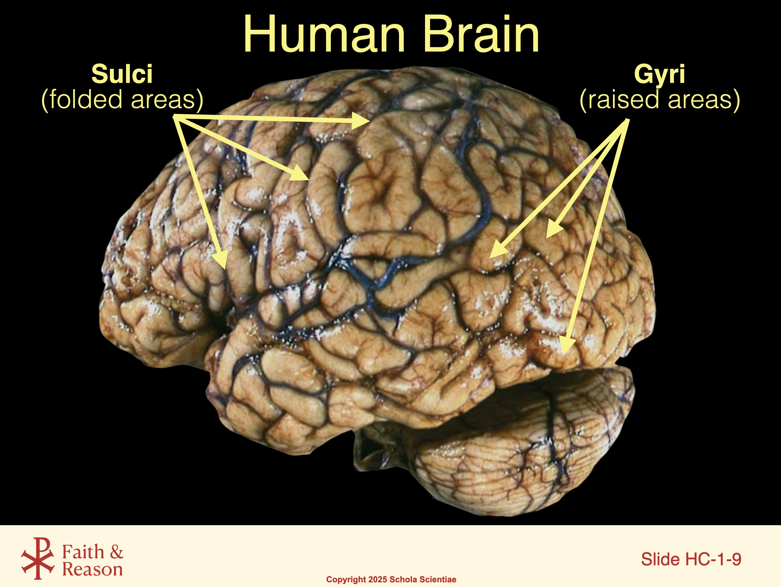
SLIDE HC-1-9
Title: Human Brain – Sulci and Gyri
Take a close look at this real human brain. You’ll notice that it’s not smooth—it’s full of ridges and grooves. These folds give the brain its familiar wrinkled look.
The raised ridges are called gyri (pronounced JY-rye).
The deep grooves are called sulci (pronounced SUL-sigh).
Why does your brain have all these folds? Because they increase the brain’s surface area, allowing more neurons to fit inside your skull. More neurons = more processing power.
Even though the brain is compact, it has the complexity and capacity to manage your body, your thoughts, your emotions, and your choices—all thanks to this folded design.
Discussion Questions and Answers:
Q1: What are gyri and sulci?
A1: Gyri are the raised folds of the brain, and sulci are the grooves or dips between them.
Q2: Why is the brain wrinkled?
A2: The folds allow more surface area to fit in a small space, which means more neurons and more brainpower.
Q3: What’s the advantage of having all those folds?
A3: They increase the number of connections the brain can make—more folds mean more thinking, memory, and control abilities.
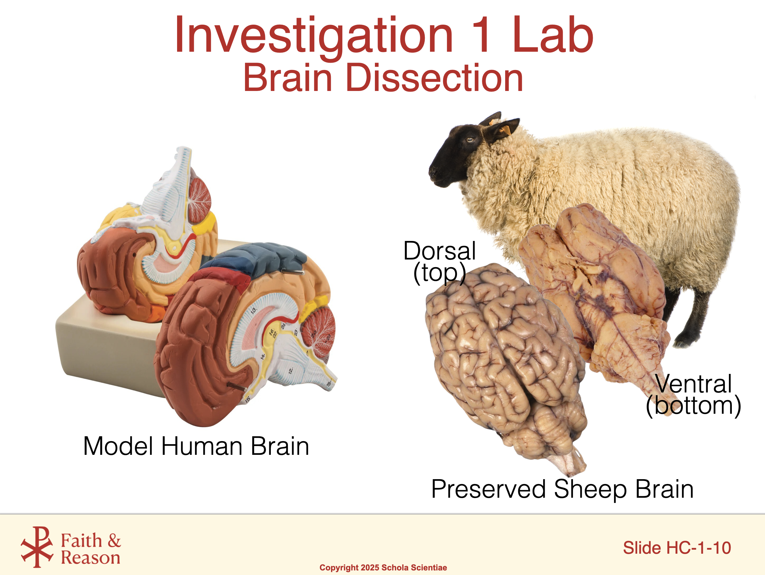
SLIDE HC-1-10
Title: Investigation 1 Lab – Brain Dissection
In this lab, we’ll get an up-close look at the real anatomy of the brain by examining a preserved sheep brain. You’ll also compare it to a model of the human brain so you can match what you see in the specimen to what you’ve learned in class.
The sheep brain may be smaller, but its structure is surprisingly similar to the human brain. You’ll be able to identify key parts like the cerebrum, cerebellum, and brainstem.
We’ll also introduce the terms ventral (meaning the bottom or underside) and dorsal (meaning the top or backside), which help us describe orientation in brain anatomy.
This lab gives you a rare chance to explore a real brain with your own eyes—and maybe even your hands. Be respectful, be curious, and take your time making observations.
Discussion Questions and Answers:
Q1: What kind of brain will we be dissecting today?
A1: A preserved sheep brain, which is similar in structure to the human brain.
Q2: Why are we using a sheep brain instead of a human one?
A2: It’s safer and more accessible, but still shares many important features with the human brain.
Q3: What do “ventral” and “dorsal” mean?
A3: “Ventral” means the bottom side, and “dorsal” means the top or backside—these terms help us describe directions in the brain.
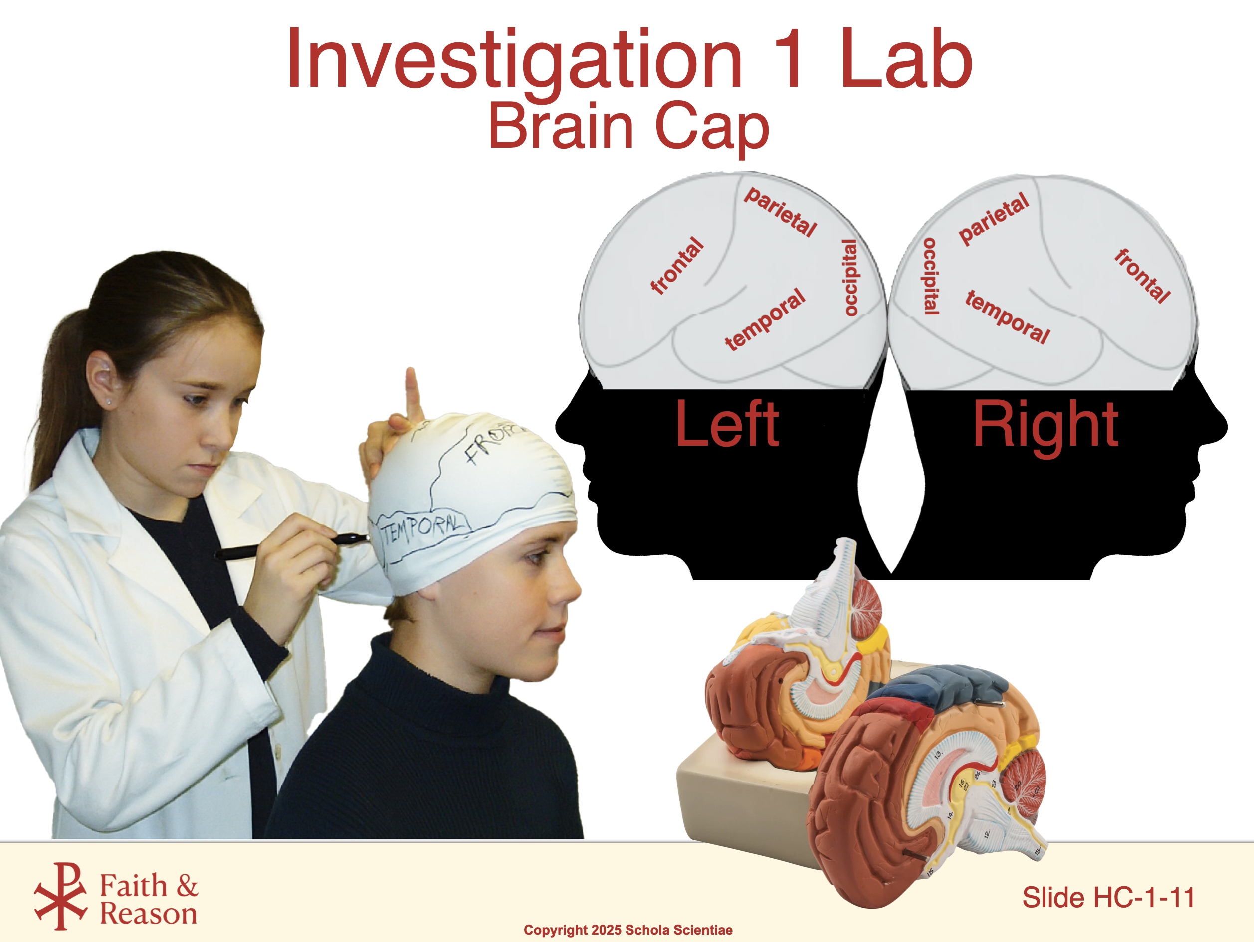
SLIDE HC-1-11
Title: Brain Cap Activity
In this activity, you’ll be labeling and mapping the major parts of the brain—on your own head!
Using a flexible “brain cap,” you’ll identify where the frontal lobe, parietal lobe, temporal lobe, and occipital lobe are located on your skull. You’ll also mark key areas like the motor cortex and sensory cortex.
Why do this? Because it helps you connect brain structure to your real experience. You’ll start to recognize which parts of your brain are involved in speaking, seeing, moving your hands, solving problems, and more.
It’s also a fun and memorable way to reinforce everything we’ve discussed about brain structure and localization of function.
Discussion Questions and Answers:
Q1: What will we be doing in the brain cap activity?
A1: We’ll map and label the different lobes of the brain on our own heads using a wearable brain cap.
Q2: What’s the point of putting brain regions on our heads?
A2: It helps us understand where different brain functions happen and connects classroom learning to our real bodies.
Q3: What are some brain parts we’ll label during this activity?
A3: The frontal, parietal, temporal, and occipital lobes, plus areas like the motor and sensory cortices.