
Teacher Portal
Investigation 1: PreLab
Get Focused:
Think about the following questions as we work through today’s PreLab.
By the end of this investigation, you should be able to answer each one confidently.
1. What role do chromosomes play in human development?
Chromosomes carry the DNA instructions that guide how every cell functions, divides, and develops. Understanding chromosomes helps explain how a single fertilized egg can grow into a complete human being.
2. Why do humans have 23 pairs of chromosomes, and why does this matter for growth?
Each pair includes one chromosome from each parent. These pairs ensure that every new cell formed during growth receives the full set of genetic instructions—critical for healthy development.
3. How does mitosis allow a tiny embryo to grow into a full-term baby?
Mitosis produces genetically identical cells again and again. This constant cell division increases cell number, allowing tissues, organs, and the entire body to grow throughout prenatal development.
4. What do the different stages of mitosis reveal about how cells divide accurately?
Each stage—prophase, metaphase, anaphase, and telophase—ensures chromosomes are copied and separated correctly. Accurate division is essential so that every new cell receives exactly 46 chromosomes.
5. Why are chromosome mistakes during early development so serious?
If a new cell receives too many or too few chromosomes, development can be disrupted. Understanding mitosis helps explain why correct chromosome number is vital for life and why errors can have major consequences.
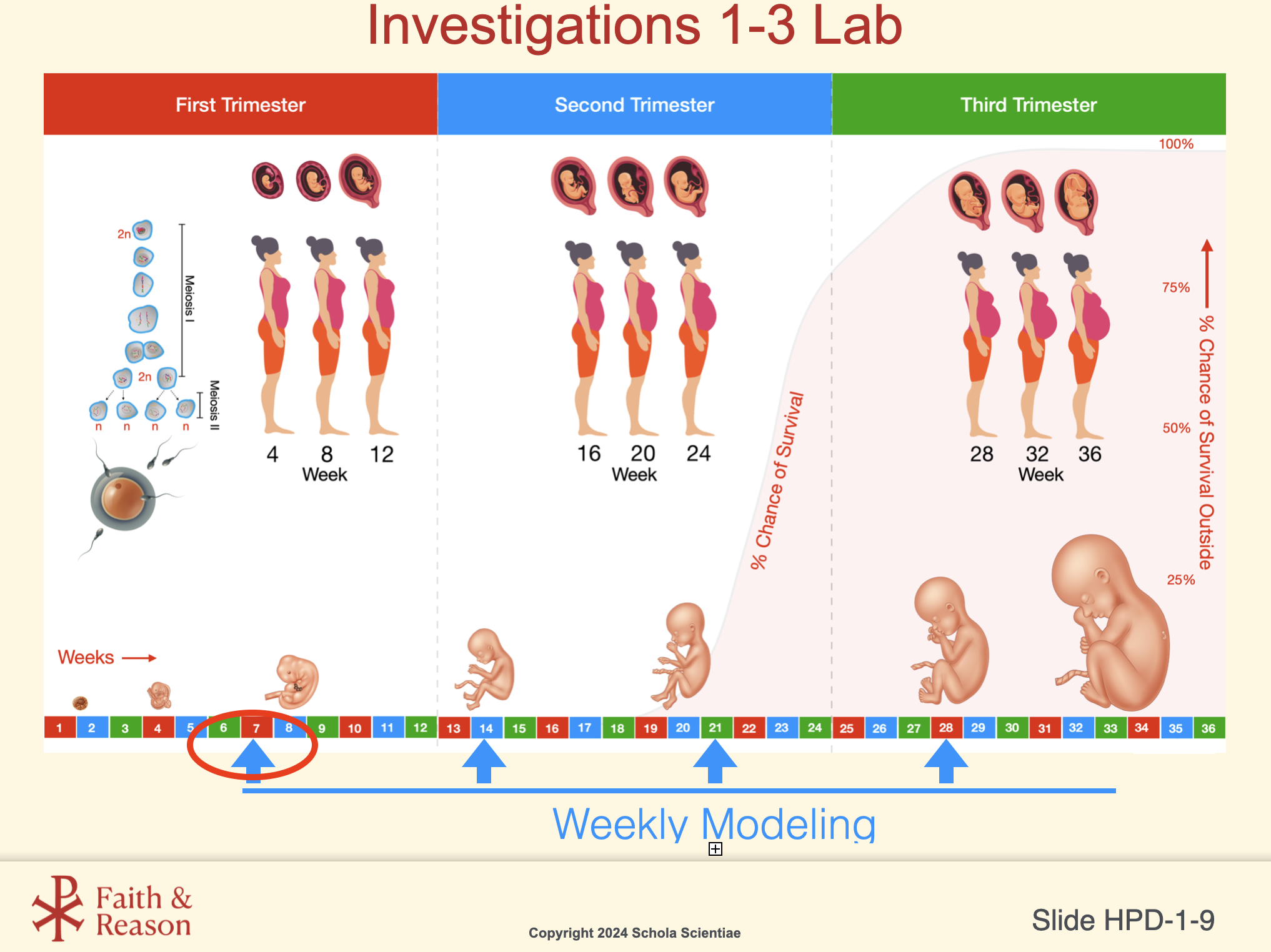
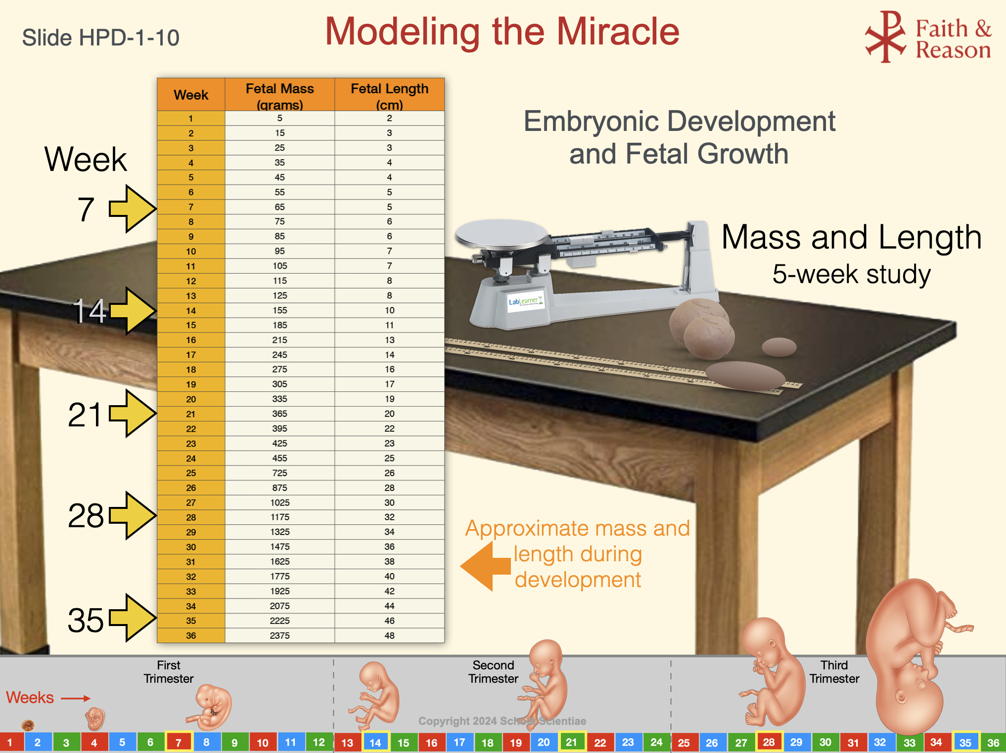
5. How does weekly fetal growth (mass and length) relate to what cells are doing?
Behind every increase in fetal size is massive cell production through mitosis. Growth charts and modeling activities help students see how cell division underlies measurable developmental changes.
Preparing for Success:
Before beginning this Investigation, students should review several key ideas from the Background Information and Concept Day slides. These ideas directly influence their success in the Mitosis Modeling Lab.
To help students succeed:
Direct them to the specific Background Readings linked below.
Encourage them to click the slide thumbnails to view important Concept Day visuals.
Review the short explanations that tell them why each concept matters for today’s investigation.
This structure removes guesswork and helps both teachers and students feel confident and prepared. These ideas directly shape how students will perform during the modeling of mitosis, chromosome number, and growth.
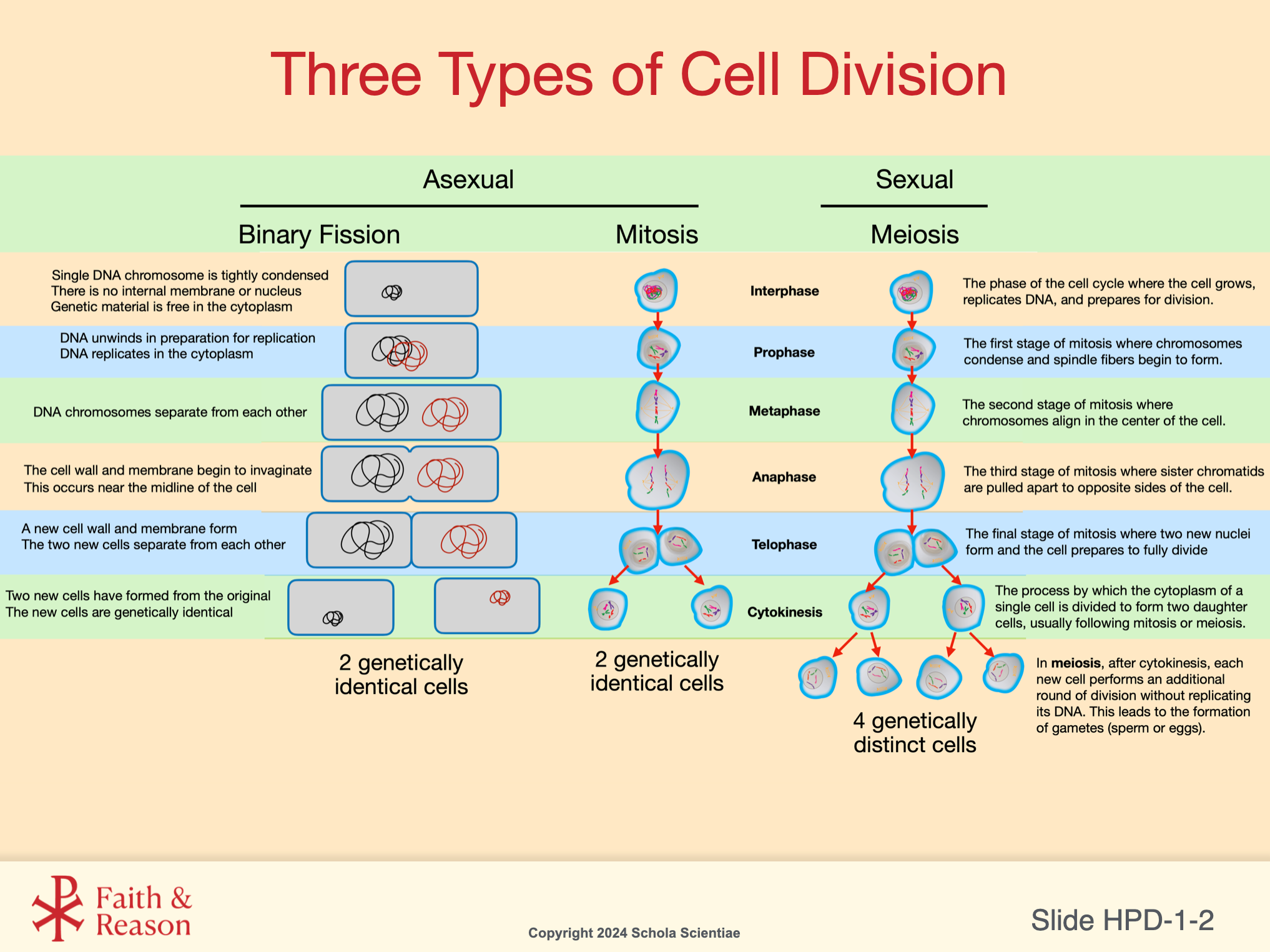
1. Chromosomes Carry Instructions for Development
Key Idea:
- Chromosomes are made of DNA, which carries the instructions for how every cell functions, divides, and develops.
Background Reading (Readings open in a new window):
Relevant Concept Slides (Click to enlarge):
Why this matters:
Students will be modeling chromosome behavior using blocks or manipulatives. They must understand what chromosomes actually represent (bundles of DNA instructions) before they attempt to model changes in chromosome number.
2. Humans Have 23 Chromosome Pairs — and This Matters for Growth
Key Idea:
Humans Have 23 Chromosome Pairs — and This Matters for Growth
Background Reading (Readings open in a new window):
Relevant Concept Slides (Click to enlarge):
Why this matters:
When students model mitosis, they must begin with the correct number of chromosomes. Miscounting the starting chromosome number is a common student mistake and leads to incorrect daughter cells later.
3. Mitosis Produces Genetically Identical Cells
Key Idea:
Mitosis ensures that each new cell receives a full set of DNA instructions—critical for growth, repair, and prenatal development.
Background Reading (Readings open in a new window):
Relevant Concept Slides (Click to enlarge):
Why this matters:
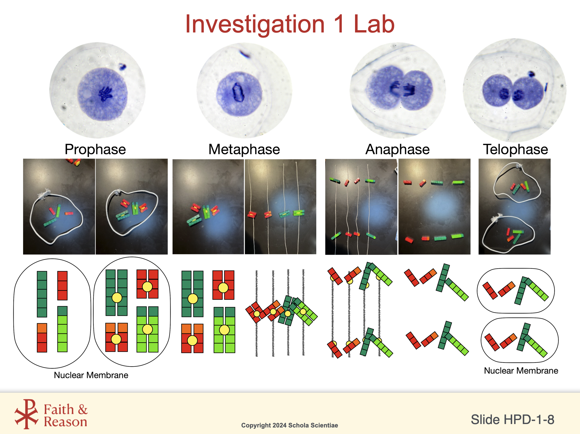
Students will model the four main stages (prophase → metaphase → anaphase → telophase).
Understanding the purpose of each stage helps them correctly manipulate their models and recognize errors in chromosome separation.
4. Mitosis Drives Massive Increases in Cell Number During Prenatal Development
Key Idea:
A human newborn has ~1.25 trillion cells. That number grows from one fertilized egg through countless rounds of mitosis.
Background Reading (Readings open in a new window):
Relevant Concept Slides (Click to enlarge):
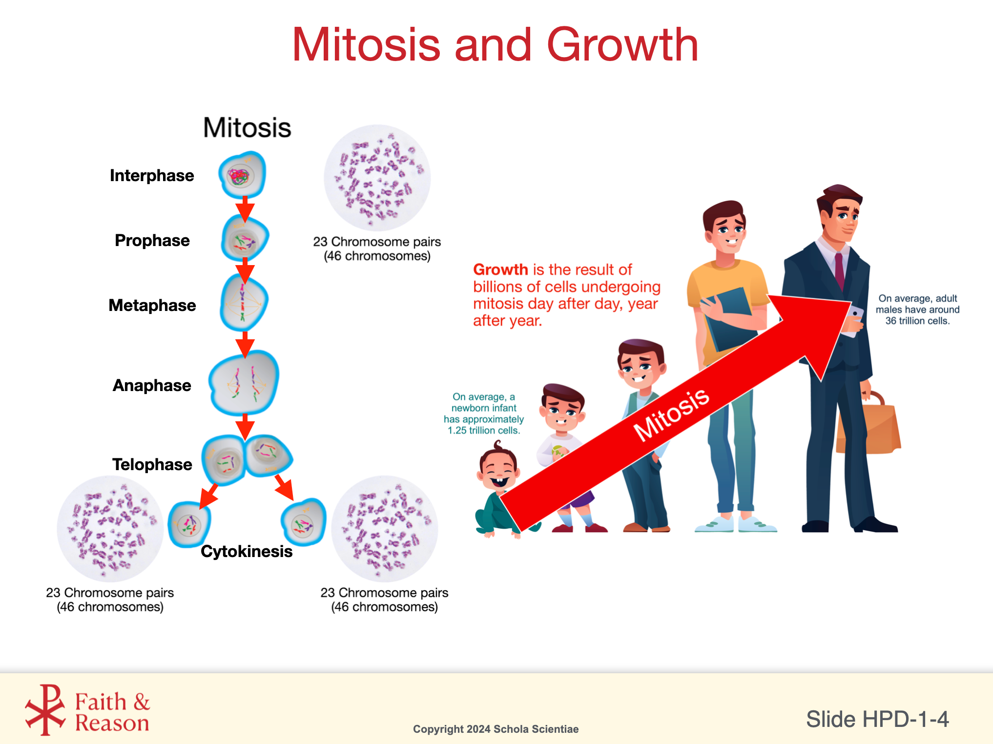
Why this matters:
Students will connect the rate of mitosis to fetal mass and length using the Modeling the Miracle chart. Understanding that growth = cell division helps them interpret lab results and trimester modeling.
5. The Stages of Mitosis Must Occur Accurately
Key Idea:
If chromosomes do not separate correctly, the resulting cells may have too many or too few chromosomes.
Background Reading (Readings open in a new window):
Relevant Concept Slides (Click to enlarge):
Why this matters:
When students perform the Modeling the Miracle activity, they will model correct chromosome separation.
This reinforces why accuracy in mitosis is essential for healthy development.
6. Growth Patterns Relate to Trimester Development
Key Idea:
Fetal growth follows predictable patterns. Understanding trimester-based changes helps students interpret the Modeling the Miracle data.
Relevant Concept Slides (Click to enlarge):
7. Students Will Apply All These Concepts in Lab
Key Idea:
The PreLab is not abstract — it is a roadmap for success in the Lab.
Students must be ready to:
Begin with the correct chromosome number.
Model chromosome duplication accurately.
Model the stages of mitosis in correct order.
Interpret fetal mass/length data as outcomes of mitotic cell increase.
Why this matters:
Students who walk into the Lab with these ideas clear will excel, make fewer construction mistakes, and produce more accurate interpretations.
Investigation Vocabulary:
Cell Division
Definition:
The process by which a cell reproduces by dividing into two new cells.
Teacher Notes — Why this matters:
This term anchors why students will model mitosis in HPD1.
Classroom Example:
Teacher asks: “Why does your body need cell division even right now?” Students connect it to repair and growth.
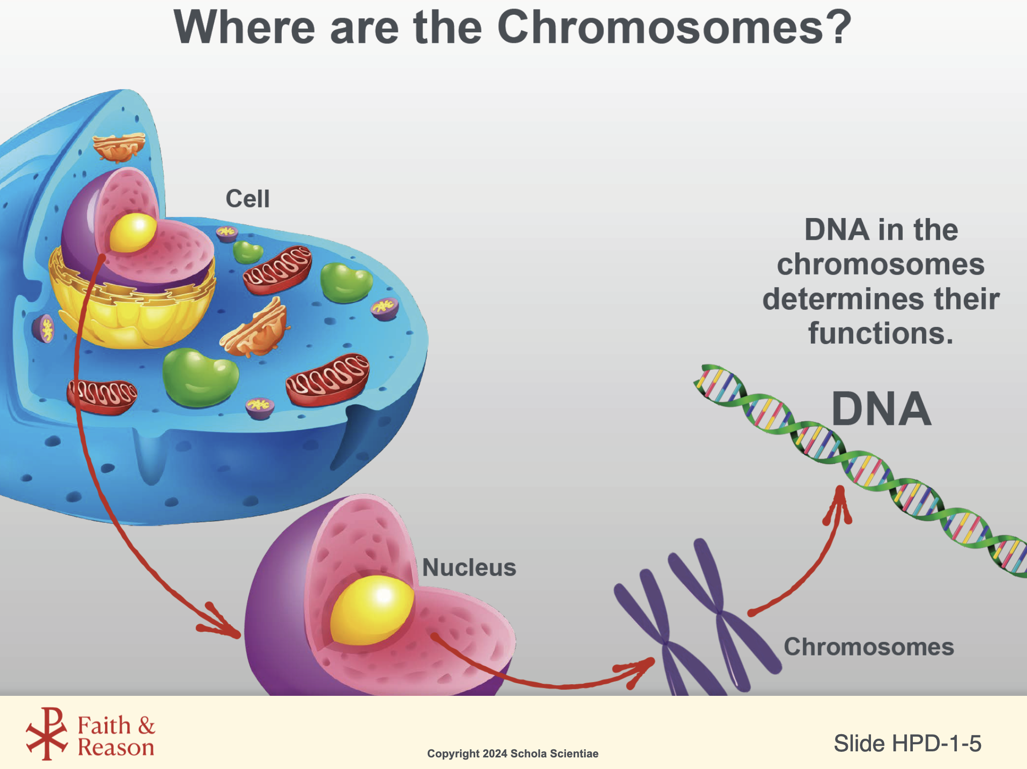
Chromosome (KROH-muh-sohm)
Definition:
A tightly coiled structure made of DNA that carries the genetic instructions for building and operating the body.
Teacher Notes — Why this matters:
Understanding chromosomes is essential for students to make sense of fertilization, cell division, genetic inheritance, and every stage of prenatal development.
Classroom Example — What this looks like:
“Every cell in your body has 46 chromosomes — the instruction sets that tell each cell what to do.”
Cytokinesis (SY-toh-kih-NEE-sis)
Definition:
The final stage of cell division in which the cytoplasm separates, forming two complete cells.
Teacher Notes — Why this matters:
Students will physically model this step using the Lab materials.
Classroom Example:
Teacher uses string to demonstrate the “pinching in” of animal cell cytokinesis.
DNA
Definition:
The molecule inside chromosomes that contains all the genetic information needed for growth, repair, and development.
Teacher Notes — Why this matters:
Students must understand DNA before they can understand how chromosomes replicate and why accurate copying is critical during mitosis.
Classroom Example — What this looks like:
“This long molecule has the instructions that make you you.”
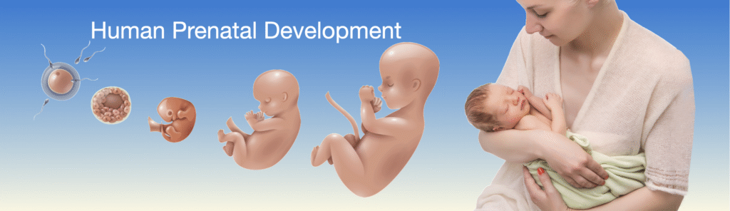
Embryo (EM-bree-oh)
Definition:
An early stage of human development before major organs are formed.
Teacher Notes — Why this matters:
This lays groundwork for understanding early prenatal growth and the weekly modeling activity.
Classroom Example:
Teacher asks students to predict: “At which week would the embryo begin looking more like a fetus?”
Fetus (FEE-tus)
Definition:
The developing human from about week 9 until birth, when major organs have begun forming.
Teacher Notes — Why this matters:
Students must distinguish embryo vs fetus to interpret the weekly fetal mass and length table.
Classroom Example:
Teacher displays Week 14 vs Week 21 models and asks: “What growth patterns do you notice?”
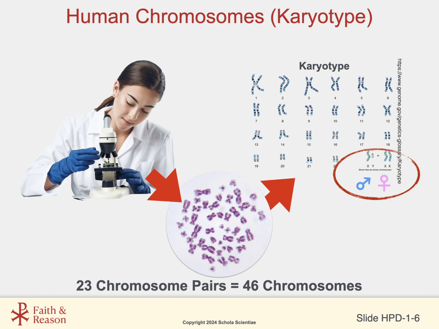
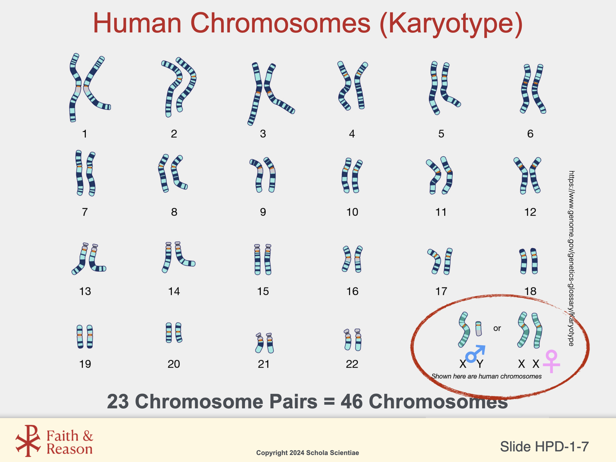
Karyotype (CARE-ee-oh-type)
Definition:
An organized image of all the chromosomes in a cell, arranged in pairs.
Teacher Notes — Why this matters:
Understanding the normal set of 23 pairs sets up later lessons on meiosis and inheritance.
Classroom Example:
Teacher shows two karyotypes and asks: “Which one is male and how do you know?”
Mitosis (my-TOE-sis)
Definition:
The process in which a single cell divides to form two genetically identical cells.
Teacher Notes — Why this matters:
Growth, tissue repair, and cell replacement all depend on mitosis. Students must understand how chromosomes copy and separate accurately.
Classroom Example:
During a review, teacher asks: “Show me—what happens in metaphase?” Students point to chromosomes lining up in the center.
Nucleus (NOO-klee-us)
Definition:
The membrane-bound structure in the cell that houses the chromosomes.
Teacher Notes — Why this matters:
Students often forget that mitosis is division of the nucleus, not the entire cell.
Classroom Example:
Teacher taps a diagram: “Where is mitosis actually happening?” Students point to the nucleus.